
|
|
|
Peripheral Nervous System- Afferent Division (Somatic) |
|
Content Peripheral Nervous System
Sensory Receptors
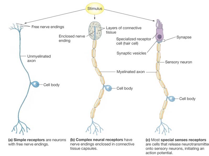
Receptors Classified by Stimulus
Receptors Classified by Stimulus Type
Stimuli exist in a variety of energy forms or modalities – heat, light, sound, pressure, chemical etc. |
|
Special Senses--External Stimuli
|
 |
Somatic Senses—Internal Stimuli
|
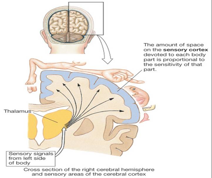 |
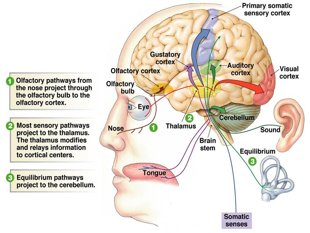
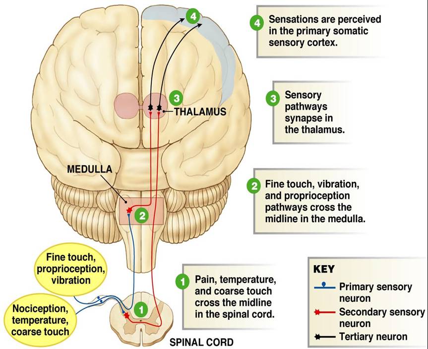
Somatic Pathways
|
 |
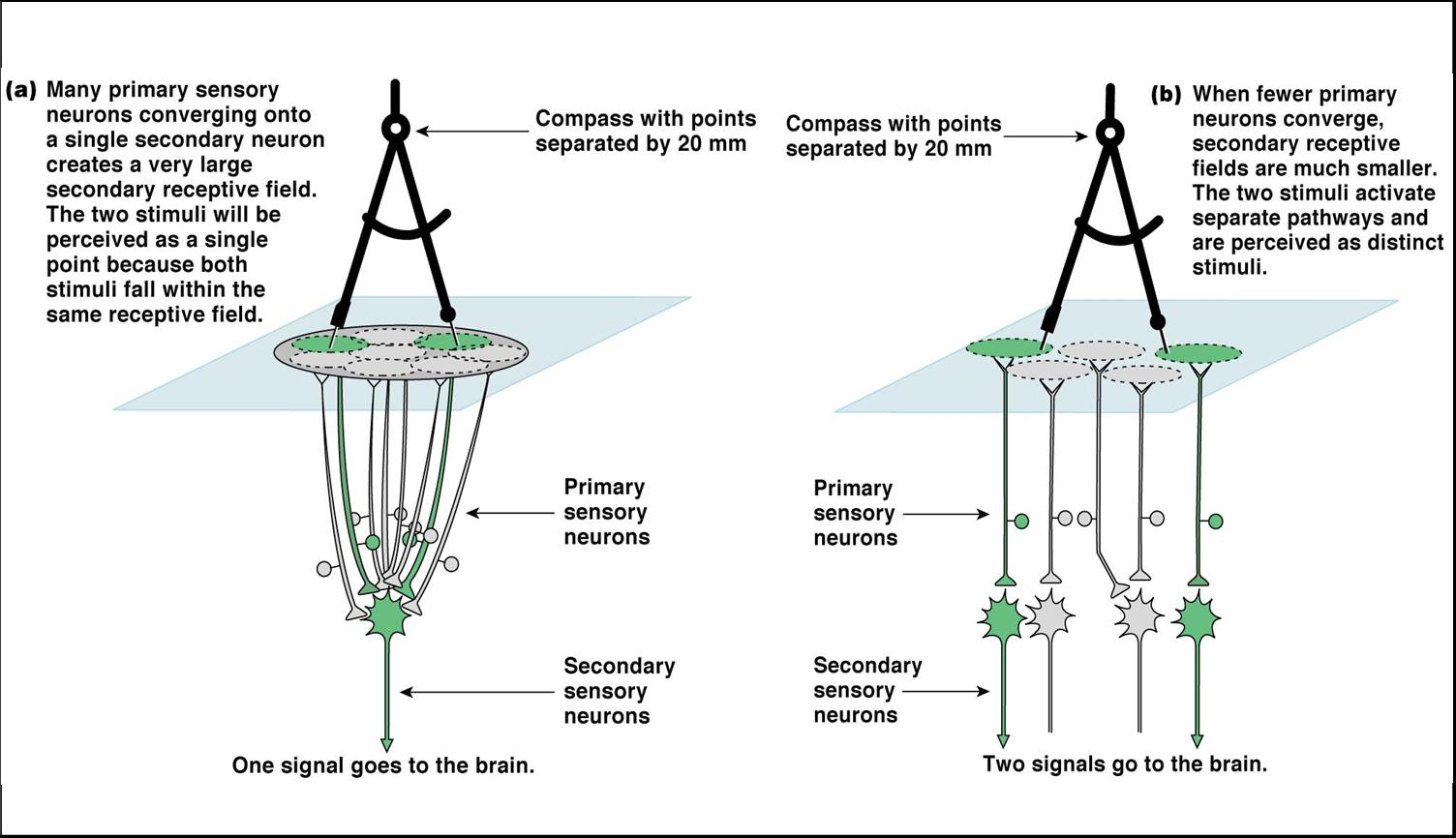
*Receptive field = area within which a receptor can detect a stimulus
Lateral inhibition; to facilitate localization and sharpen contrast, the most strongly activated pathway at the center inhibits the less excited pathways from the fringe areas
Adaptation occurs when sensory receptors are subjected to an unchanging stimulus
|
|
- Tonic receptors do not adapt at all or very slowly, important when maintaining information about a stimulus is valuable – stretch, pain receptors
- Phasic receptors – rapidly adapt, useful in situations where it is important to signal a change in stimulus – tactile (touch) receptors |
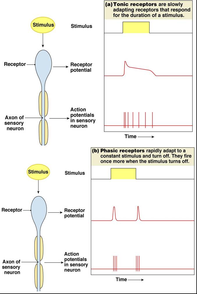 |
Adaptation mechanisms
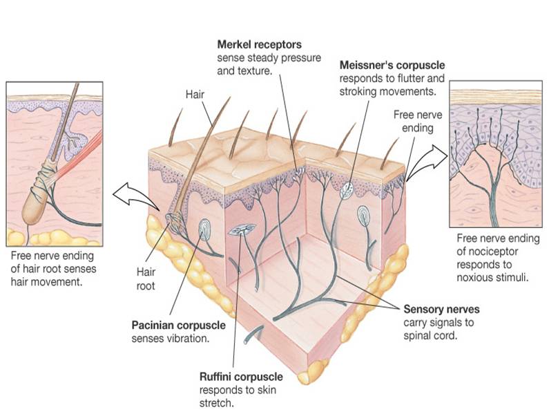
Tactile Receptors (pressure)
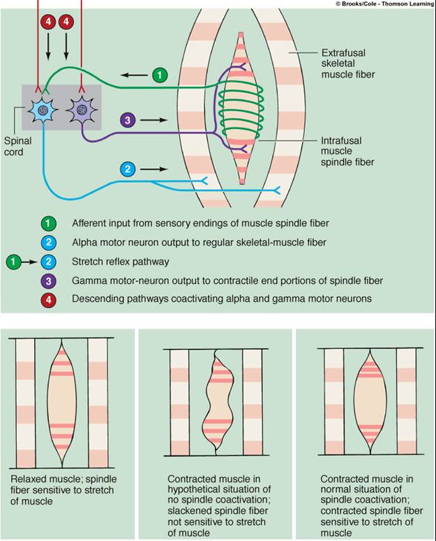
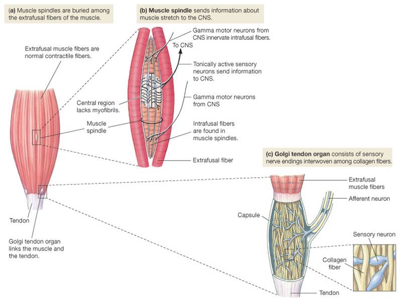
Proprioceptors - located in muscles, tendons, joints and internal ear and provide information about body position and movement.
-Tendon Organs (Golgi tendon organs) - consists of sensory fiber penetrating a thin capsule of connective tissue and entwining around a few collagen fibers, found at the junctions of a tendon with a muscle, help protect tendons and associated muscles from damage due to excessive tension or stretching
Classic example is patellar tendon, or knee-jerk reflex
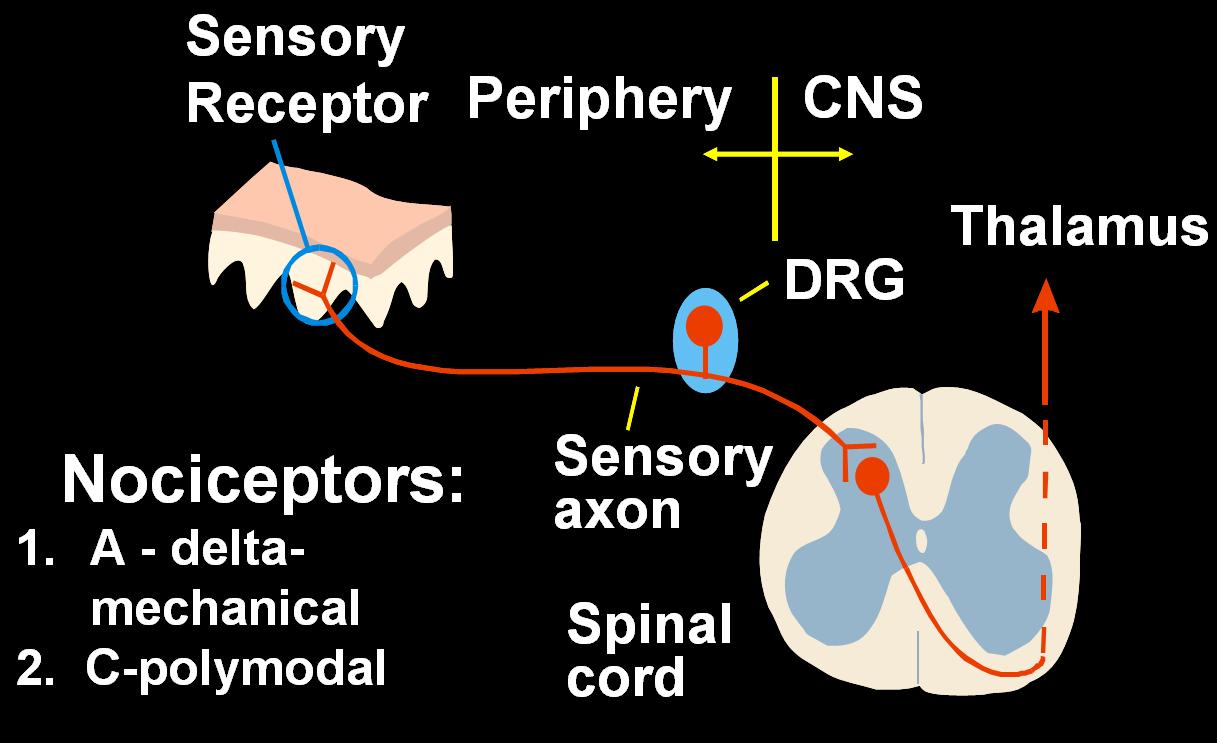
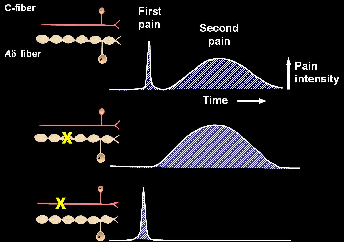
Mechanical (crushing, cutting, pinching) and thermal (extreme temperatures) are transmitted over small myelinated A-delta fibers – 30m/sec fast pain pathway Polymodal repond to all kinds of damaging stimuli and is carried by small unmyelinated C-fibers 12m/sec slow pain pathway
Processing of Pain - Afferent pathway
|
|
This material is based upon work supported by the Nursing, Allied Health and Other Health-related Educational Grant Program, a grant program funded with proceeds of the State’s Tobacco Lawsuit Settlement and administered by the Texas Higher Education Coordinating Board. |