Content
Branches of the Peripheral Efferent Division
Autonomic Branch
Parasympathetic
Sympathetic
Subdivisions of Autonomic System
Alpha Receptors
Beta Receptors
Somatic Nervous System
Motor Unit
Neuromuscular Junction
| The efferent division of the peripheral nervous system provides a communication link between the central nervous system (brain and spinal cord) and the activities of muscle and glands – the effector organs |
Branches of the Peripheral Efferent Division
- Autonomic Branch – involuntary cardiac, smooth muscle, exocrine and some endocrine glands
- Somatic Branch– voluntary movement and secretions
Autonomic Branch
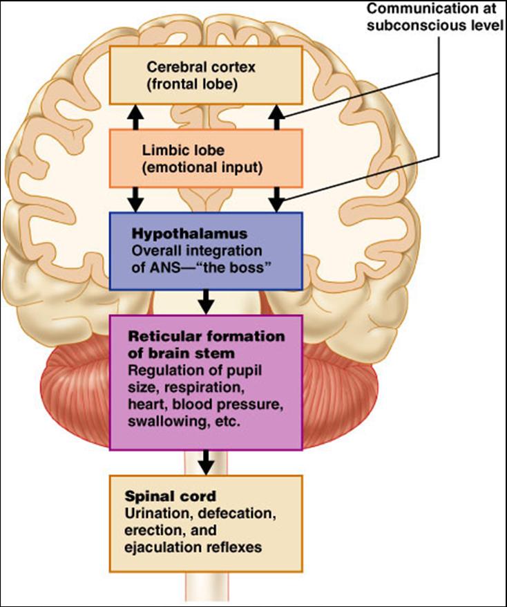
The ANS operates without conscious control, it is primarily regulated by the hypothalamus and the medulla oblongata with input from the limbic system and other regions of the cerebrum.
 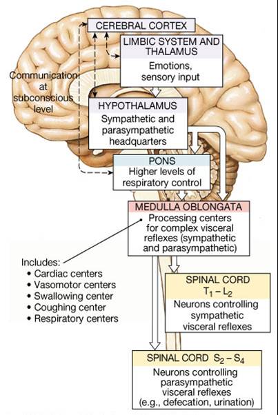
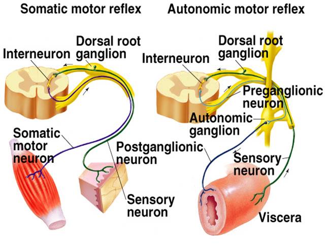
The afferent component of the ANS consists of general visceral sensory neurons. Interoreceptors such as chemoreceptors (CO2 levels) and mechanoreceptors (degree of stretch of organs and vessels).
Afferent signals are not consciously recognized unless intense enough to cause pain or nausea from damaged viscera, fullness of bladder, angina pectoris (inadequate blood flow to heart)
The efferent component consists of the autonomic motor neurons that excite or inhibit visceral activities of effector tissues; cardiac muscle, smooth muscle, glands
Efferent responses include automatic activities beyond conscious control - dilation/constriction of pupils, accommodation of lens, dilation of blood vessels, heartbeat, GI tract movement, glandular secretions.
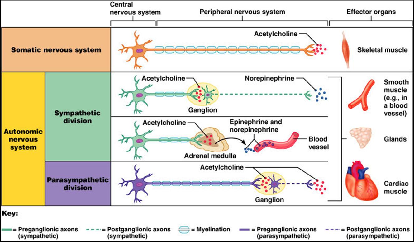
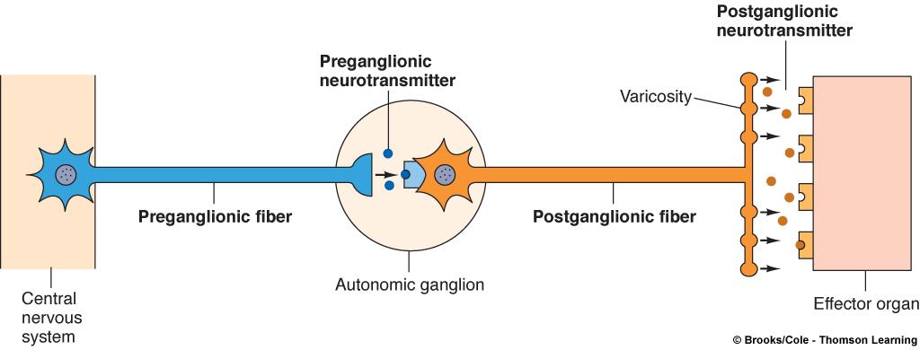
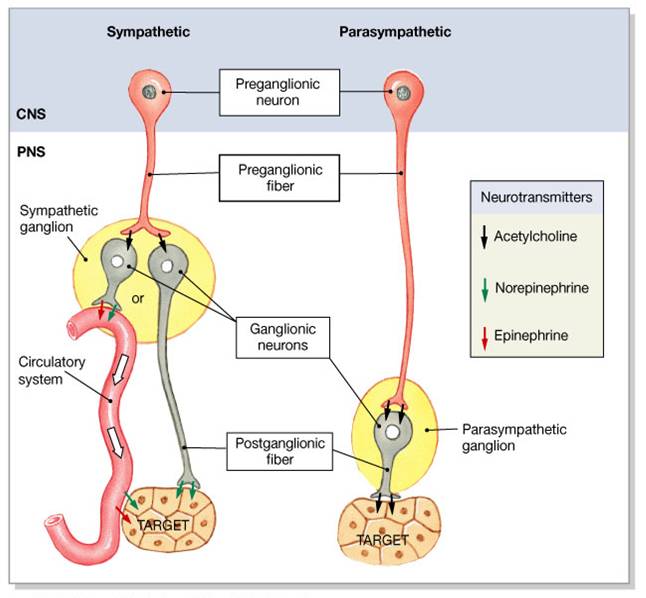
Autonomic pathway consists of a two neuron chain:
The first motor neuron called the preganglionic neuron, has its cell body in the CNS and its myelinated axon called the preganglionic fiber extends to autonomic ganglion.
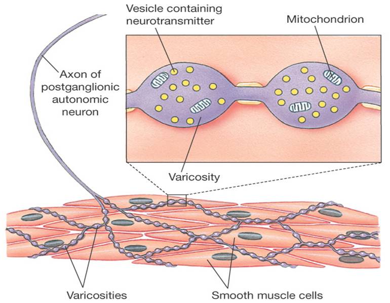
The second motor neuron called the postganglionic neuron has its soma in that autonomic ganglion and its unmyelinated axon called the postganglionic fiber extends directly to the effector cell or organ
Two Subdivisions of Autonomic System
- Sympathetic
- Parasympathetic
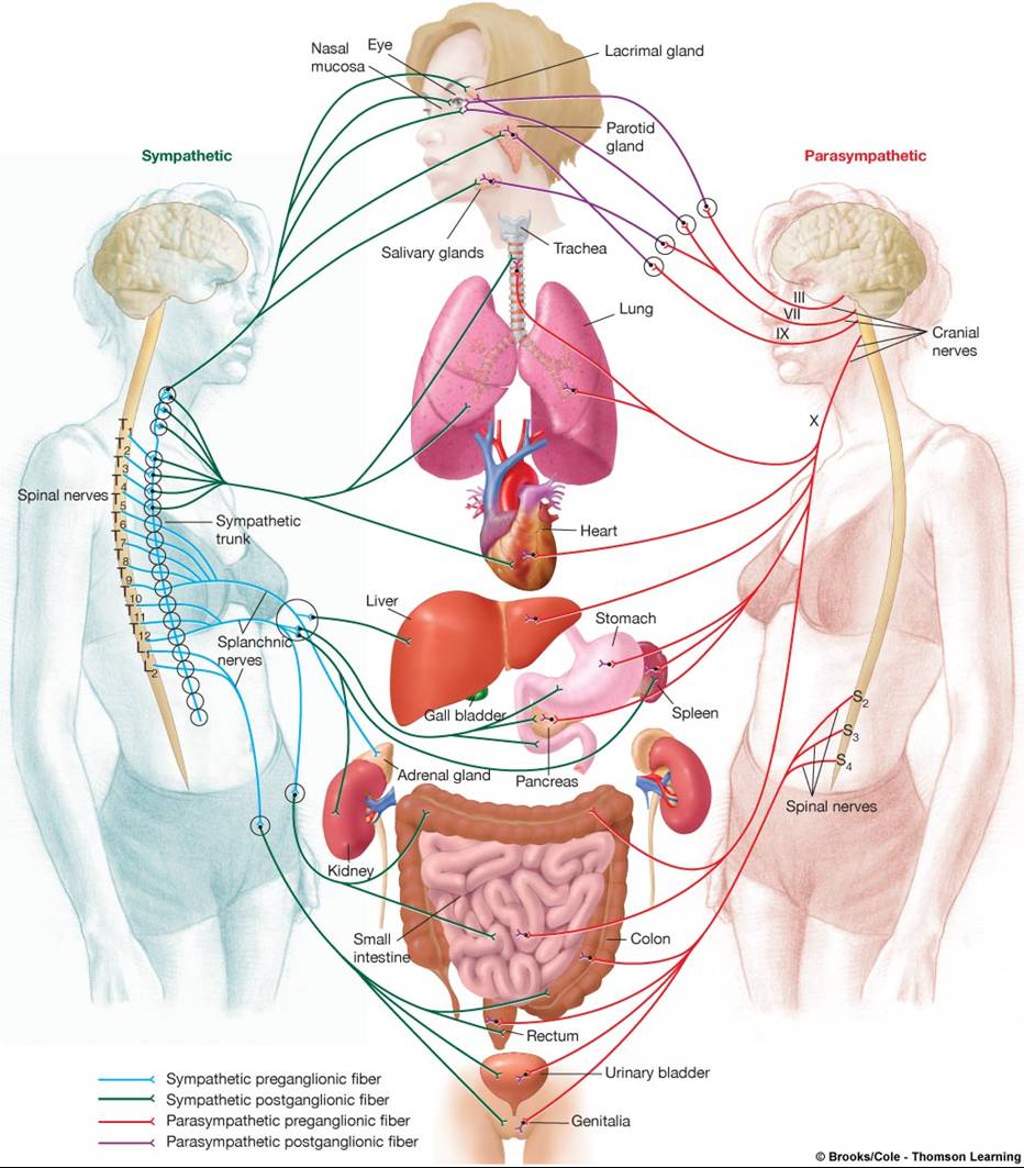
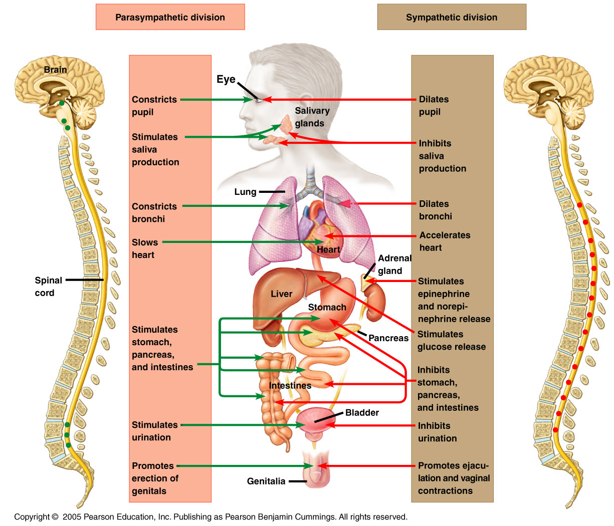
Parasympathetic
- Concerned with keeping body energy use as low as possible, and SLUDD: salivation, lacrimation, urination digestion, defecation
- Parasympathetic preganglionic neuron have cell body in the nuclei of the oculomotor, facial, glossopharyngeal, and vagus cranial nerves and in the lateral gray horns of the second through fourth sacral segments. Their axons pass to terminal ganglia near or within the visceral receptor.
- Parasympathetic postganglionic neurons cell bodies are found in the terminal ganglia and their axons synapse with single visceral effectors.
Sympathetic
- “fight or flight” system, excites body in emergency or threatening situations
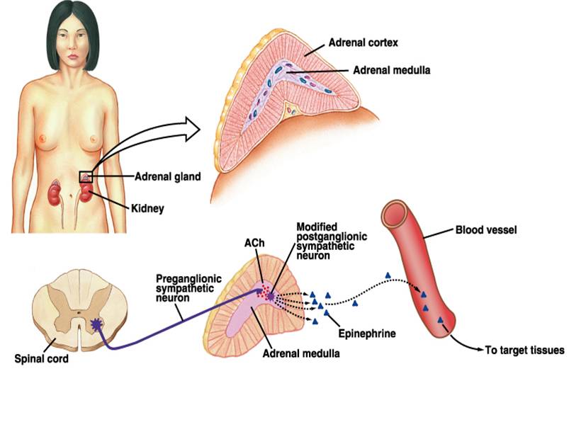
- Sympathetic preganglionic neurons have cell bodies in the lateral gray horns of the 12 thoracic segments and the first 2-3 lumbar segments and their axons synapse in the ganglia of the sympathetic trunk (paravertebral ganglia) located in parallel rows on both sides of the vertebral column or prevertebral ganglia which are located close to large abdominal cavities
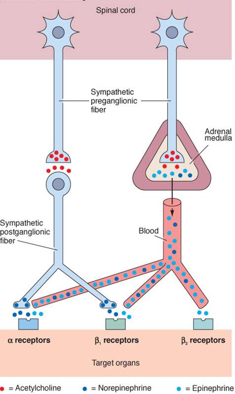
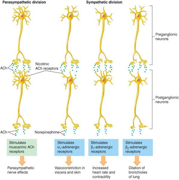
Only 2 Neurotransmitters Are Involved: Acetylcholine, Norepinephrine (Noradrenaline)
Acetylcholine released by cholinergic neurons are used by
1. All sympathetic preganglionic neurons
2. All parasympathetic preganglionic neurons
3. All parasympathetic postganglionic neurons
4. Some sympathetic postganglionic neurons
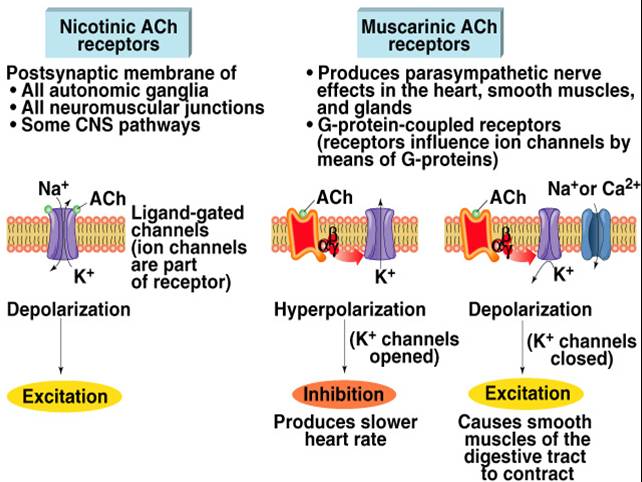
Acetylcholine released from both sympathetic and parasympathetic preganglionic fibers binds to cholinergic receptors:
Nicotinic receptors are found on the postganglionic cell bodies in all autonomic ganglia and cause depolarization by opening both Na+ and K+ channels
Muscarinic receptors are found on smooth, cardiac muscle and glands. They bind with acetylcholine released from parasympathetic postganglionic fibers and are linked to G proteins that activate 2nd messenger systems that lead to the target cell response
 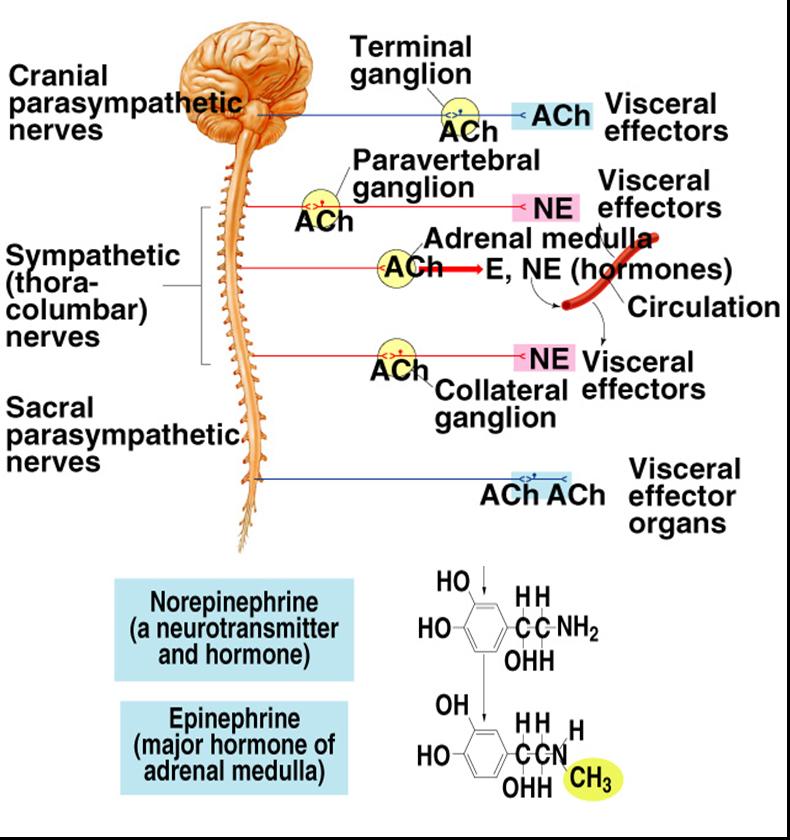
Epinephrine/Norepinephrine (adrenalin) released by adrenergic neurons, mostly sympathetic postganglionic fibers
Alpha Receptors Sensitive to Norepinephrine
Alpha-1 receptors elicit response from Ca+ second messenger system an excitatory response, present in most sympathetic target tissues
Ex. Vasoconstriction due to contraction of smooth muscle
Alpha-2 blocks cAMP production an inhibitory response,
Ex. Decreased smooth muscle contraction in digestive tract
Beta Receptors
Beta-1 sensitive to both norepinephrine and epinephrine cause excitatory response
Ex. Primarily found in heart increasing rate and force of cardiac contraction
Beta-2 sensitive to epinephrine, generally inhibitory
Ex. vasodilation caused by relaxation of smooth muscle
Beta-1 and -2, both receptors bring about target cell response through cAMP 2nd messenger system

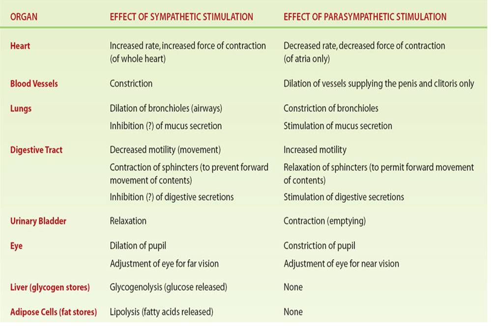
Effects of Parasympathetic and Sympathetic Divisions on Various Organs
Somatic Nervous System
Consists of the axons of motor neurons which originate in the spinal cord and terminate on skeletal muscle
Acetylcholine released from a motor neuron stimulates muscle contraction
Motor neurons are the final common pathway by which various regions of the CNS exert control over skeletal muscle activity
The areas of the CNS that influence skeletal muscle activity by acting through the motor neurons are the spinal cord, motor regions of the cortex, basal nuclei, cerebellum, and brain stem
Motor Unit
Consists of a motor neuron and the muscle fibers it stimulates. A single motor unit may contain from 2 to 2000 muscle fibers
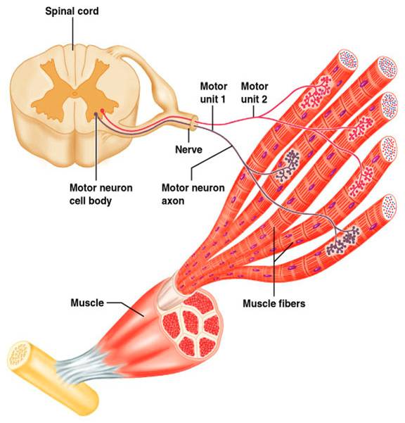
A motor neuron transmits a nerve impulse (action potential) to a neuromuscular junction.
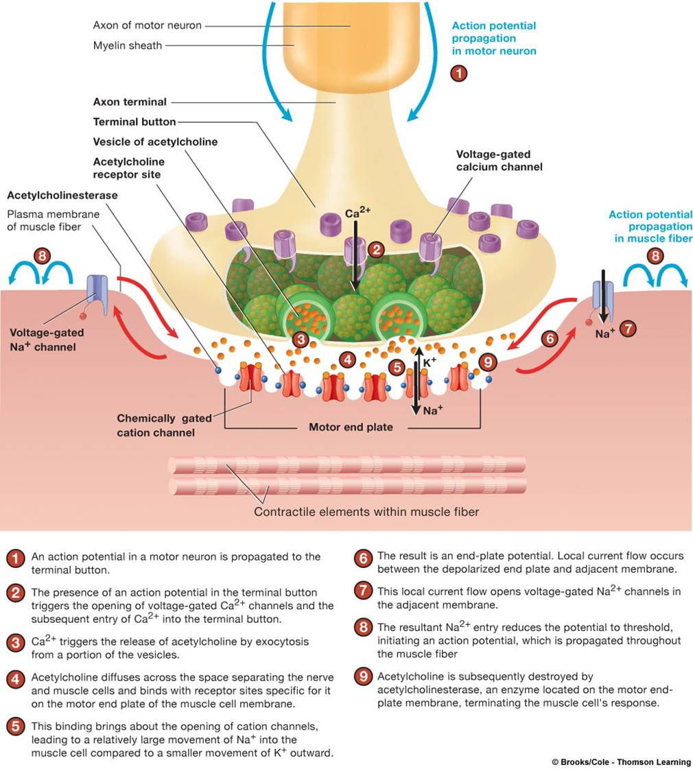
Neuromuscular Junction
Is the r egion where the of the motor neuron axon terminal and muscle fiber motor end plate make contact and communicate (synapse) .
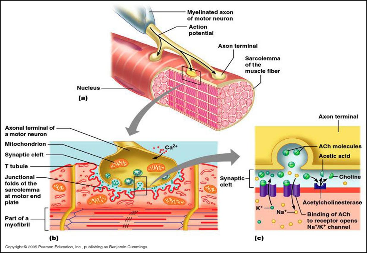
Voltage-regulated calcium channels in the axon termincal open and allow Ca2+ to enter the axon
Ca2+ inside the axon terminal causes some of the synaptic vesicles to fuse with the axon membrane and release ACh into the synaptic cleft (exocytosis)
The synaptic end bulbs releases acetylcholine from the synaptic vesicles which diffuses across the synaptic cleft (gap) to the motor end plate and attaches to ACh receptors, binding of ACh to receptors on the sarcolemma which is polarized - there is a potential difference (voltage) across the membrane.
When Ach binds to its receptors on the motor end plate, chemically (ligand) gated ion channels in the receptors open and allow Na+ and K+ to move across the membrane, resulting in a transient change in membrane potential - depolarization
End plate potential
End plate potential is a local depolarization that creates and spreads an action potential across the sarcolemma, stronger than an EPSP due to more NT, greater surface area of membrane, greater number of receptors
Top ...... Main Page |