Urinary System
Content
Functions of the Urinary System
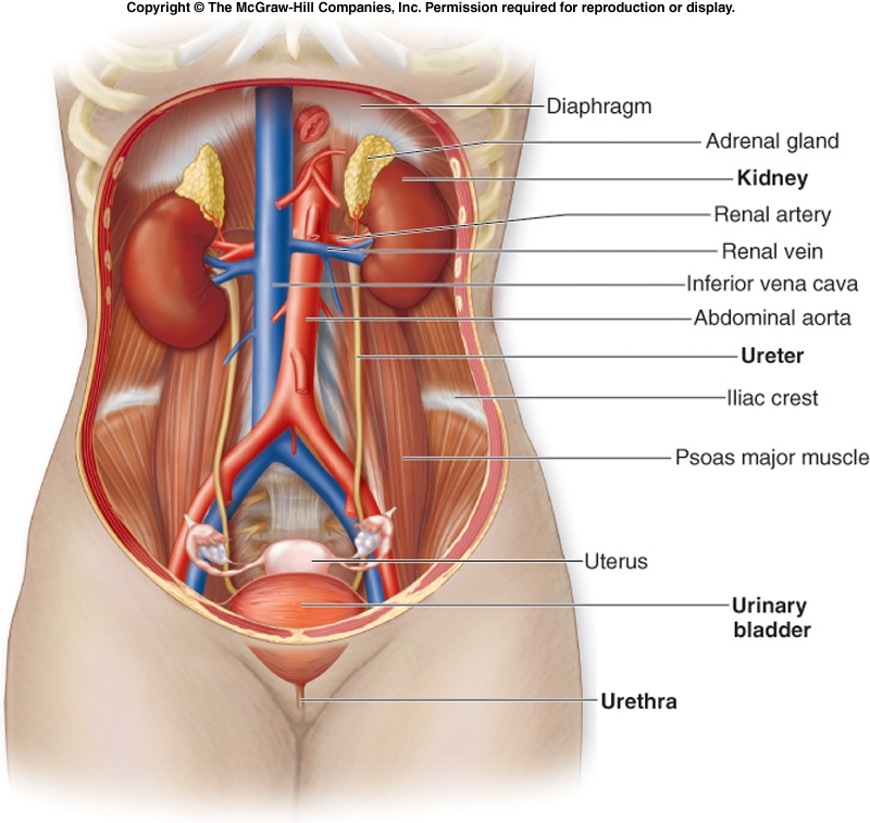
Renal Anatomy
Blood Supply to the Kidneys
Nephrons
Glomerular Filtration Rate (GFR)
Extrinsic Controls
Renin-Angiotensin Mechanism
Reabsorption: Secondary Active Transport
Tubular Secretion
Urine Formation
Atrial Natriuretic Peptide (ANP) Activity
Control of Urine Volume
Loop of Henle: Countercurrent Multiplication
Renal & Creatinine Clearance
|
Functions of the Urinary System
- Filters Waste Products from Blood
- Excretion of water and sodium chloride (NaCl) is regulated in conjunction with cardiovascular, endocrine, & central nervous system
-The urinary system eliminates in the urine different waste products such as ammonia and urea (both formed when amino acids are broken down), uric acid (formed when nucleic acids are broken down), creatinine (from muscles), end products of hemoglobin metabolism, hormone metabolites, and foreign substances (e.g., drugs, pesticides, & other chemicals ingested in the food)
-The blood is filtered by the kidney through 3 processes called filtration, reabsorption, and secretion. The wastes leave the body as urine.
- Conserves Valuable Nutrients
The urinary system ensures glucose, amino acids and other valuable nutrients are not lost from the urine
- Regulates Ion Levels in the Plasma
The urinary system regulates ion (electrolyte) levels in the plasma by regulating the amount of sodium, potassium, chloride and other ions lost in the urine.
- Regulates Blood pH
The urinary system regulates blood pH by regulating the number of H+ and bicarbonate ions (HCO3-) lost in the urine.
The k idneys work in concert with lungs to regulate the pH in a narrow limits of buffers within body fluids.
Regulates Blood Volume
The urinary system regulates blood volume by:
1) releasing renin, a hormone that after a series of reactions eventually restricts salt and water loss at the kidneys.
2) adjusting the volume of water lost in the urine
- Regulates RBC Production
If oxygen levels in the blood are low, the kidneys release erythropoietin, a hormone that stimulates the hemocytoblasts (stem cells in the bone marrow) to increase red blood cell formation. Having more RBCs allows the blood to transport more oxygen.
- Stores Urine
The bladder stores the urine until it is convenient to excrete it.
- Excretes Urine
The urethra transports urine from the urinary bladder to the outside of the body.
- Produces and secretes hormones:
- Renin activates the renin-angiotensin-aldosterone system, thus regulating blood pressure & Na+,K+ balance
- Prostaglandins/kinins bradykinin = vasoactive, leading to modulation of renal blood flow & along with angiotensin II affect the systemic blood flow
- Erythropoietin stimulates red blood cell formation by bone marrow
|
|
| |
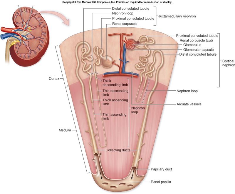
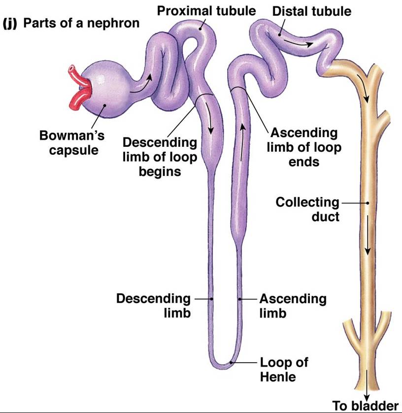
Renal Anatomy
The functional units of the kidneys are called nephrons. Each kidney is made up of millions of nephrons.
Each nephron is made up of the following:
- Glomerulus
- Bowman’s Capsule
- Glomerular Capillaries
- Proximal Convoluted Tubule (PCT)
- Loop of Henle
- Distal Convoluted Tubule (DCT)
- Collecting Duct
Click here for an animation that reviews the microanatomy of the kidney.
|
 |
 |
 |
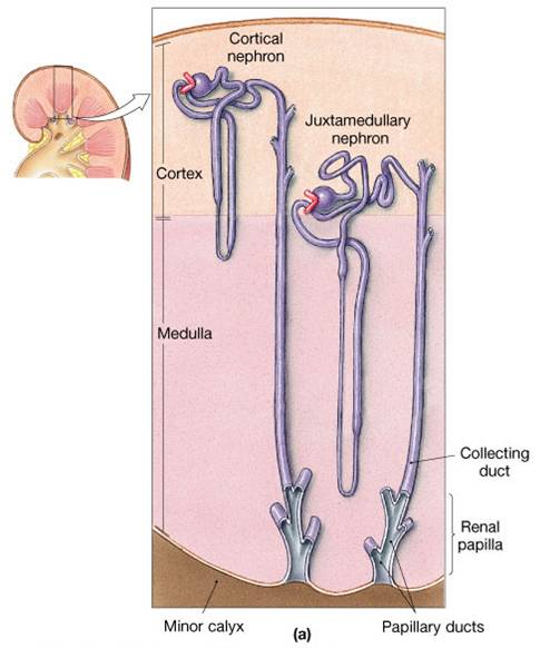
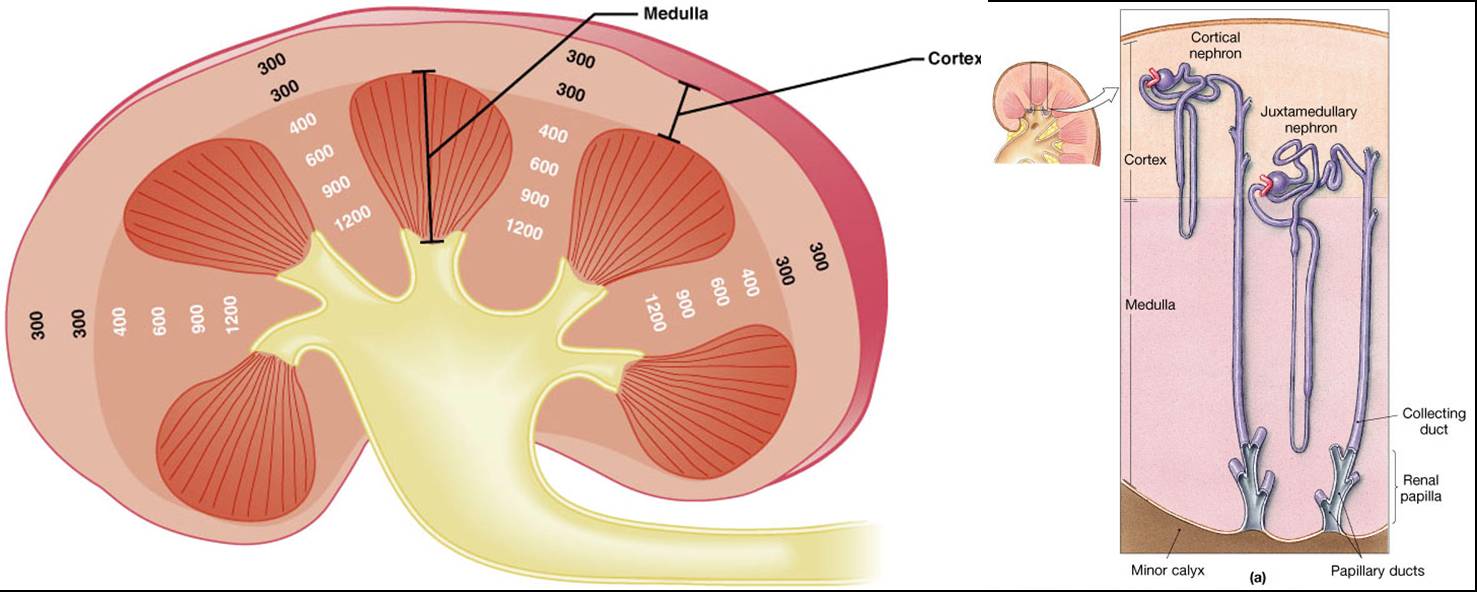
Two Types of Nephrons:
- -~85% of all nephrons
- -Are located in the cortex
- Juxtamedullary nephrons,
- -Are closer (juxta = next to) the renal medulla
- -The loops of Henle extend deep into the renal pyramids
|
 |
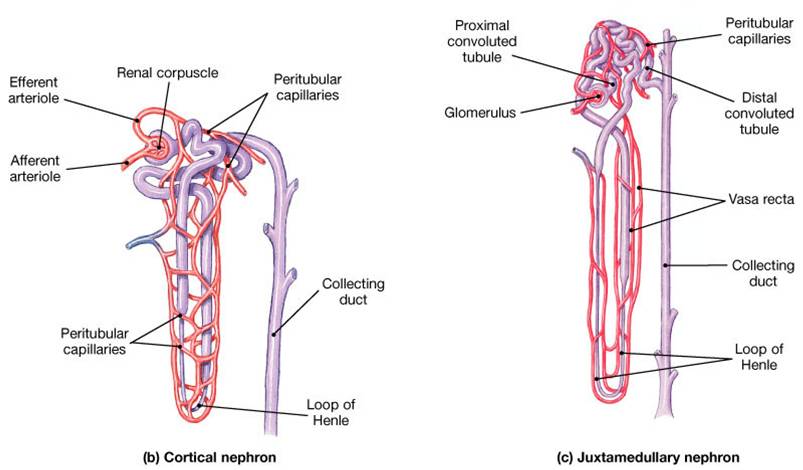
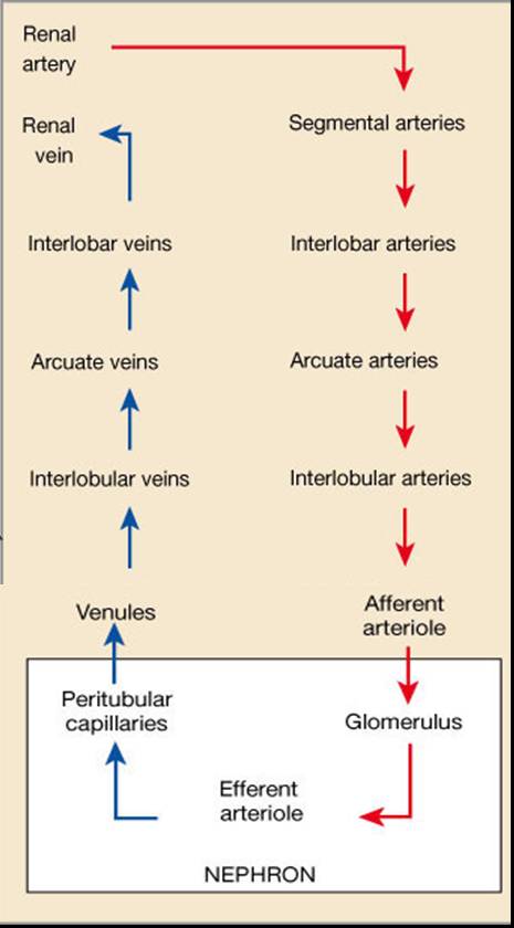
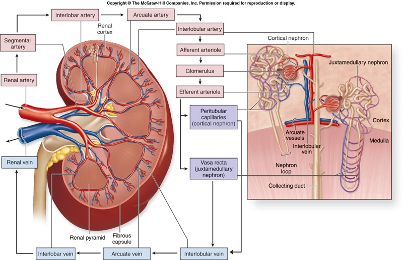
Blood Supply to the Kidneys
- Blood travels from the afferent arteriole to a ball of capillaries in the nephron called a glomerulus
- Blood leaves the nephron via the efferent arteriole
- Blood travels from efferent arteriole to the peritubular capillaries and vasa recta
|
 |
 |
|
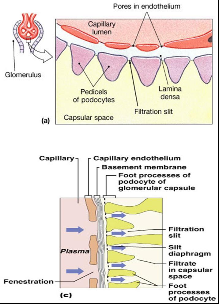
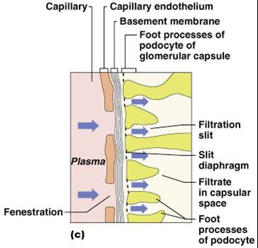
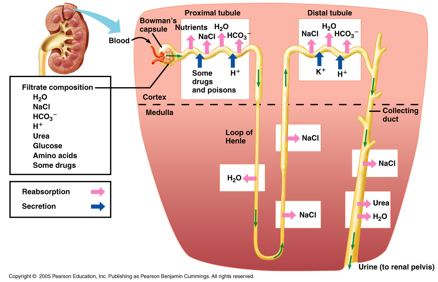
Filtrate Composition
- Glomerular filtrate is produced from blood plasma
- The filtrate m ust pass through:
- Pores between endothelial cells of the glomerular capillary
- Basement membrane - Acellular gelatinous membrane made of collagen and glycoprotein
- Filtration slits formed by podocytes
- Filtrate is similar to plasma in terms of concentrations of salts and of organic molecules (e.g., glucose, amino acids) except it is essentially protein-free
- Glomerular filtration barrier restricts the filtration of molecules on the basis of size and electrical charge
- Neutral solutes:
- Solutes smaller than 180 nanometers in radius are freely filtered
- Solutes greater than 360 nanometers are not
- Solutes between 180 and 360 nm are filtered to various degrees
- Serum albumin is anionic and has a 355 nm radius, only ~7 g is filtered per day (out of ~70 kg/day passing through glomeruli)
- In a number of glomerular diseases, the negative charge on various barriers for filtration is lost due to immunologic damage and inflammation, resulting in proteinuria (i.e.increased filtration of serum proteins that are mostly negatively charged).
- The principles of fluid dynamics that account for tissue fluid in the capillary beds apply to the glomerulus as well
- Filtration is driven by Starling forces across the glomerular capillaries. Changes in these forces and in renal plasma flow alter the glomerular filtration rate (GFR)
- The glomerulus is more efficient than other capillary beds because:
- -Its filtration membrane is significantly more permeable
- -Glomerular blood pressure is higher
- -It has a higher net filtration pressure
- -Plasma proteins are not filtered and are used to maintain oncotic (colloid osmotic) pressure of the blood
|
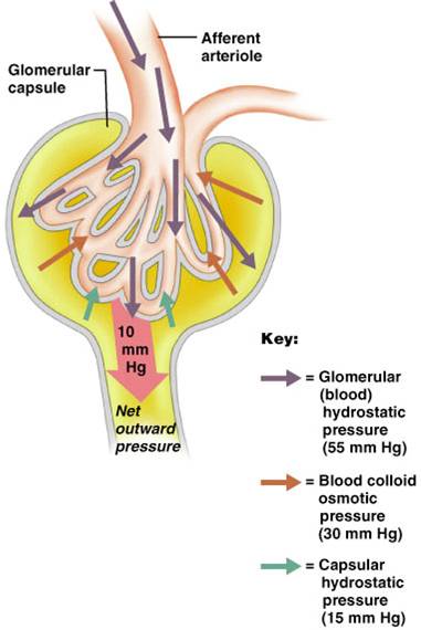
Forces Involved in Glomerular Filtration
- Net Filtration Pressure (NFP) This is the pressure responsible for filtrate formation
- NFP equals the glomerular hydrostatic pressure (HPg) minus the oncotic pressure of glomerular blood (OPg) plus capsular hydrostatic pressure (HPc)
NFP = HPg – (OPg + HPc)
NFP = 55 – (30 + 15) = 10
|
 |
Glomerular Filtration Rate (GFR)
The total amount of filtrate formed per minute by the kidneys
- Filtration rate factors:
- Total surface area available for filtration and membrane permeability (filtration coefficient = Kf)
- Net filtration pressure (NFP)
- GFR = Kf x NFP
- GFR is directly proportional to the NFP
- Changes in GFR normally result from changes in glomerular capillary blood pressure
Kidney’s Receive 20-25% of CO
- At NFP of 10mmHG
- Filtration fraction: ~ 20% of the plasma that enters the glomerulus is filtered
- Males = 180 L of glomerular filtrate per day - 125ml/min
- Females = 160 L per day – 115ml/min
- For 125ml/min, renal plasma flow = 625ml/min
- 55% of blood is plasma, so blood flow = 1140ml/min
- 1140 = 22% of 5 liters
- Required for adjustments and purification, not to supply kidney tissue
Regulation of Glomerular Filtration
- If the GFR is too high, needed substances cannot be reabsorbed quickly enough and are lost in the urine
- If the GFR is too low -everything is reabsorbed, including wastes that are normally disposed of
- Control of GFR normally result from adjusting glomerular capillary blood pressure
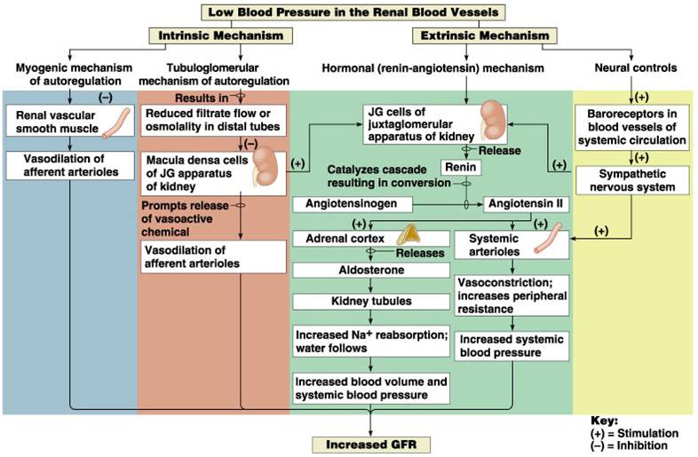
- 3 mechanisms control the GFR
- Renal autoregulation (intrinsic system)
- Neural controls
- Hormonal mechanism (the renin-angiotensin system)
Autoregulation of GFR
- Under normal conditions (MAP =80-180mmHg) renal autoregulation maintains a nearly constant glomerular filtration rate
- 2 mechanisms are in operation for autoregulation:
- Myogenic mechanism
- Arterial pressure rises, afferent arteriole stretches
- Vascular smooth muscles contract
- Arteriole resistance offsets pressure increase; RBF (& hence GFR) remain constant.
- Tubuloglomerular feed back mechanism for autoregulation:
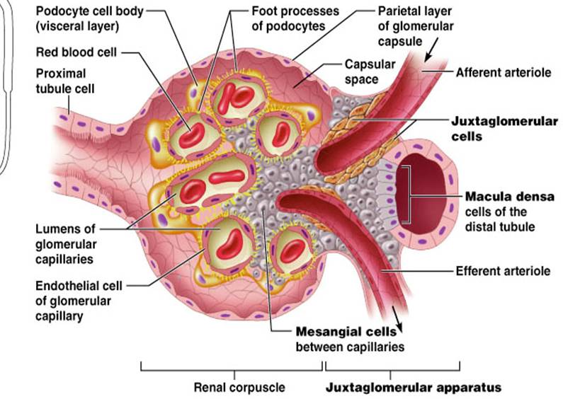
- Feedback loop consists of a flow rate (increased NaCl) sensing mechanism in macula densa of juxtaglomerular apparatus (JGA)
- Increased GFR (& RBF) triggers release of vasoactive signals
- Constricts afferent arteriole leading to a decreased GFR (& RBF)
Extrinsic Controls
- When the sympathetic nervous system is at rest:
- Renal blood vessels are maximally dilated
- Autoregulation mechanisms prevail
- Under stress:
- Norepinephrine is released by the sympathetic nervous system
- Epinephrine is released by the adrenal medulla
- Afferent arterioles constrict and filtration is inhibited
- The sympathetic nervous system also stimulates the renin-angiotensin mechanism
- A drop in filtration pressure stimulates the Juxtaglomerular apparatus (JGA) to release renin and erythropoietin
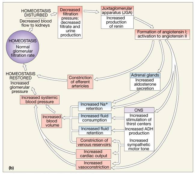
Renin-Angiotensin Mechanism
Renin release is triggered by:
- Reduced stretch of the granular JG cells
- Stimulation of the JG cells by activated macula densa cells
- Direct stimulation of the JG cells via b1-adrenergic receptors by renal nerves
- Renin acts on angiotensinogen to release angiotensin I which is converted to angiotensin II
- Angiotensin II
- Causes mean arterial pressure to rise
- Stimulates the adrenal cortex to release aldosterone
- As a result, both systemic and glomerular hydrostatic pressure rise
Other Factors Affecting Glomerular Filtration
- Prostaglandins (PGE2 and PGI2)
- Vasodilators produced in response to sympathetic stimulation and angiotensin II
- Are thought to prevent renal damage when peripheral resistance is increased
- Nitric oxide – vasodilator produced by the vascular endothelium
- Adenosine – vasoconstrictor of renal vasculature
- Endothelin – a powerful vasoconstrictor secreted by tubule cells
Control of Kf
- Mesangial cells have contractile properties, influence capillary filtration by closing some of the capillaries – effects surface area
- Podocytes change size of filtration slits
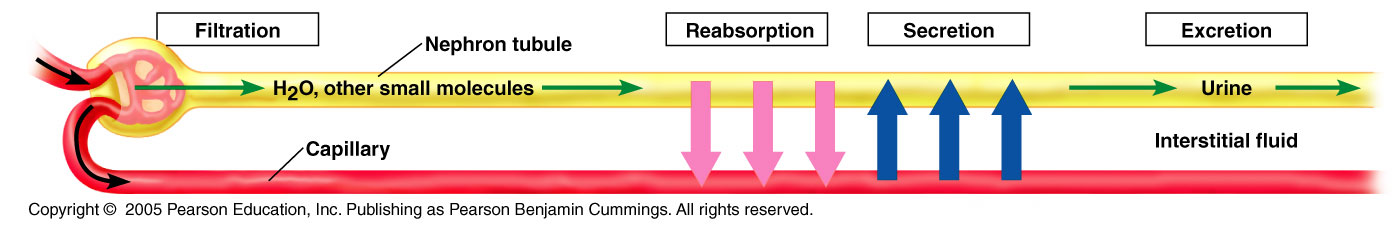
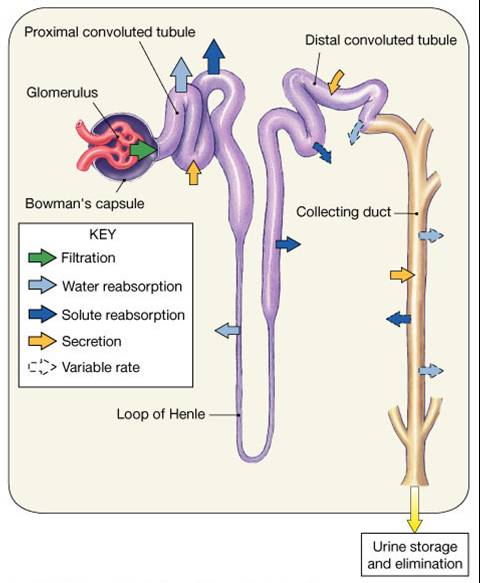
Process of Urine Formation
Urine formation depends on 3 processes:
- Glomerular filtration
- Tubular reabsorption of the substance from the tubular fluid into blood
- Tubular secretion of the substance from the blood into the tubular fluid
Click here for an animation that describes in detail the process of urine formation.
- Amount Excreted in Urine = Amount Filtered through glomeruli into renal proximal tubule MINUS amount reabsorbed into capillaries PLUS amount secreted into the tubules
1. Reabsorption and Secretion
- Accomplished via
- diffusion
- osmosis
- active and facilitated transport
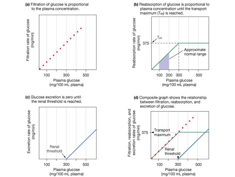
- Carrier proteins have a transport maximum (Tm) which determines renal threshold for reabsorption of substances in tubular fluid
- A transport maximum (Tm):
- Reflects the number of carriers in the renal tubules available
- Exists for nearly every substance that is actively reabsorbed
- When the carriers are saturated, excess of that substance is excreted
|
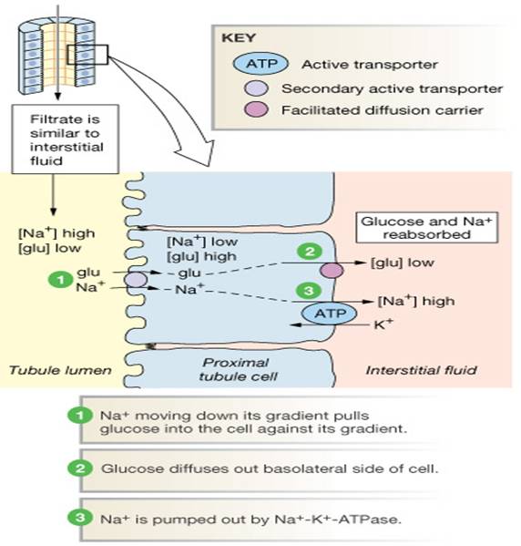
Reabsorption: Secondary Active Transport
- Na+ linked active transport
- Cotransport
- Proximal tubule, key site
|
 |
Non-Reabsorbed Substances
- Substances are not reabsorbed if they:
- Lack carriers
- Are not lipid soluble
- Are too large to pass through membrane pores
- Urea, creatine, and uric acid are the most important nonreabsorbed substances
|
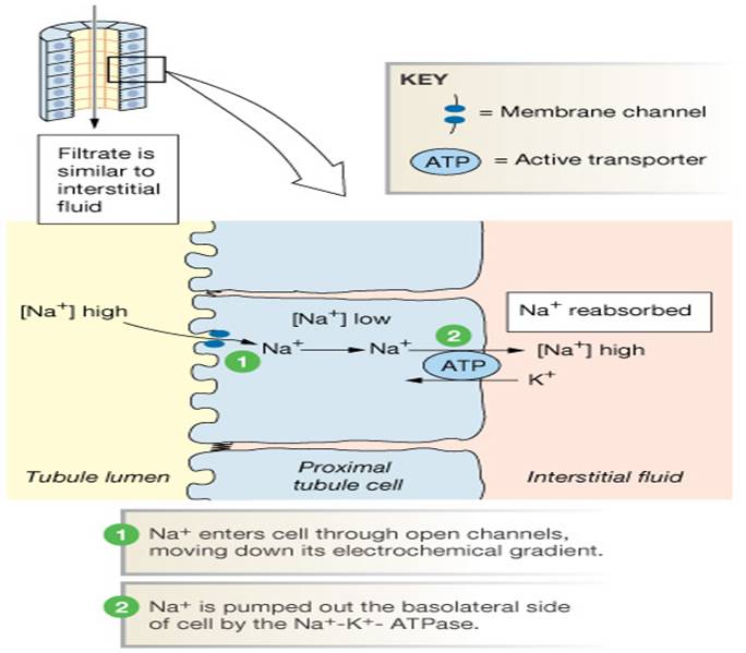
Sodium Reabsorption:
- Sodium reabsorption is almost always by active transport via a Na+-K+ ATPase pump
- Sodium reabsorption provides the energy and the means for reabsorbing most other solutes
- Water by osmosis
- Organic nutrients and selected cations by secondary (coupled) active transport
|
 |
2.Tubular Secretion
- Essentially reabsorption in reverse, where substances move from peritubular capillaries or tubule cells into filtrate
- Tubular secretion is important for:
- Disposing of substances not already in the filtrate
- Eliminating undesirable substances such as urea and uric acid
- Ridding the body of excess potassium ions
- Controlling blood pH
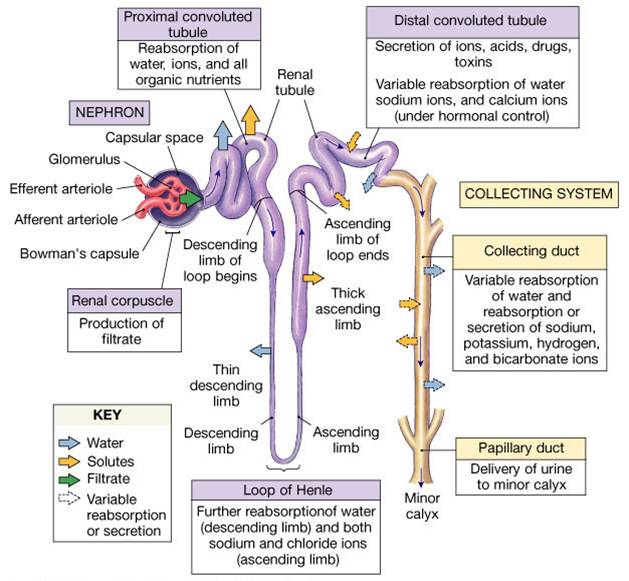
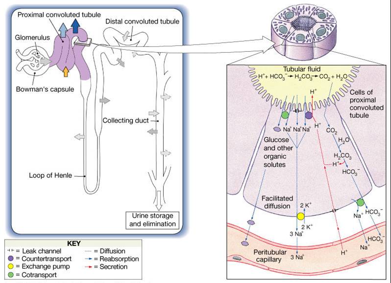
Reabsorption and Secretion at the PCT
- Glomerular filtration produces fluid similar to plasma without proteins
- The PCT reabsorbs 60-70% of the filtrate produced
- Sodium, all nutrients, cations, anions, and water
- Urea and lipid-soluble solutes
- Small proteins
- H+ secretion also occurs in the PCT
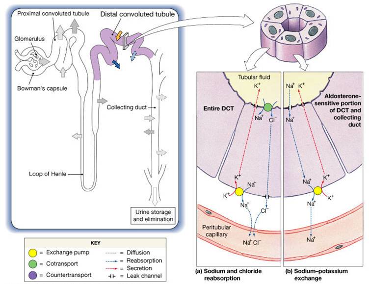
Reabsorption and Secretion at the DCT
DCT performs final adjustment of urine
- Active secretion or absorption
- Absorption of Na+ and Cl-
- Secretion of K+ and H+ based on body pH
- Water is regulated by ADH (vasopressin)
- Na+, K+ regulated by aldosterone
Atrial Natriuretic Peptide (ANP) Activity
- ANP reduces blood Na+ which:
- Decreases blood volume
- Lowers blood pressure
- ANP lowers blood Na+ by:
- Acting directly on medullary ducts to inhibit Na+ reabsorption
- Counteracting the effects of angiotensin II
- Antagonistic to aldosterone and angiotensin II.
- Promotes Na+ and H20 excretion in the urine by the kidney.
- Indirectly stimulating an increase in GFR reducing water reabsorption

|
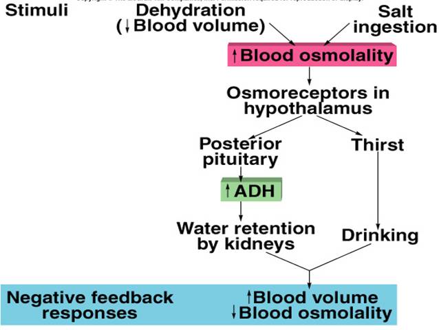
Regulation by ADH
- Released by posterior pituitary when osmoreceptors detect an increase in plasma osmolality.
- Dehydration or excess salt intake:
- Produces sensation of thirst.
- Stimulates H20 reabsorption from urine.
Click here for an animation on the release of ADH in response to decreased blood volume.
The animation is followed by practice questions. |
 |
Control of Urine Volume
- Urine volume and osmotic concentration are regulated by controlling water reabsorption
- Precise control allowed via facultative water reabsorption
Regulation of Urine Concentration and Volume
- Osmolality
- The number of solute particles dissolved in 1L of water
- Reflects the solution’s ability to cause osmosis
- Body fluids are measured in milliosmols (mOsm)
- The kidneys keep the solute load of body fluids constant at about 300 mOsm
- This is accomplished by the countercurrent mechanism
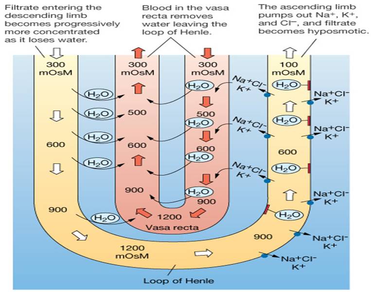
Countercurrent Mechanism
- Interaction between the flow of filtrate through the loop of Henle (countercurrent multiplier) and the flow of blood through the vasa recta blood vessels (countercurrent exchanger)
- The solute concentration in the loop of Henle ranges from 300 mOsm to 1200 mOsm
- Vasa Recta prevents loss of medullary osmotic gradient equilibrates with the interstitial fluid
- Maintains the osmotic gradient
- Delivers blood to the cells in the area
Loop of Henle: Countercurrent Multiplication
- The descending loop: relatively impermeable to solutes, highly permeable to water
- The ascending loop: permeable to solutes, impermeable to water
- Collecting ducts in the deep medullary regions are permeable to urea
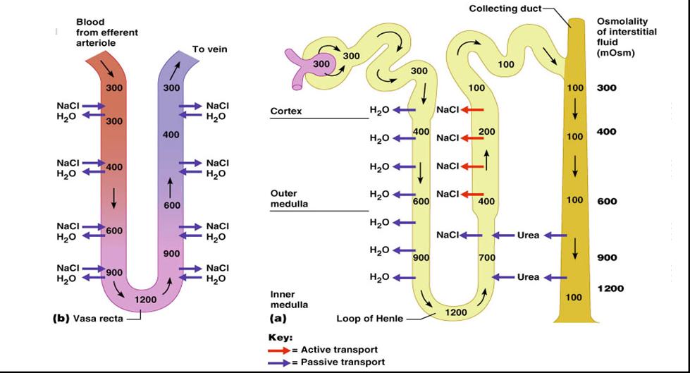
Water Reabsorption in Descending Loop of Henle
- Countercurrent multiplier and exchange
- Medullary osmotic gradient
- H2O®ECF®vasa recta vessels
|
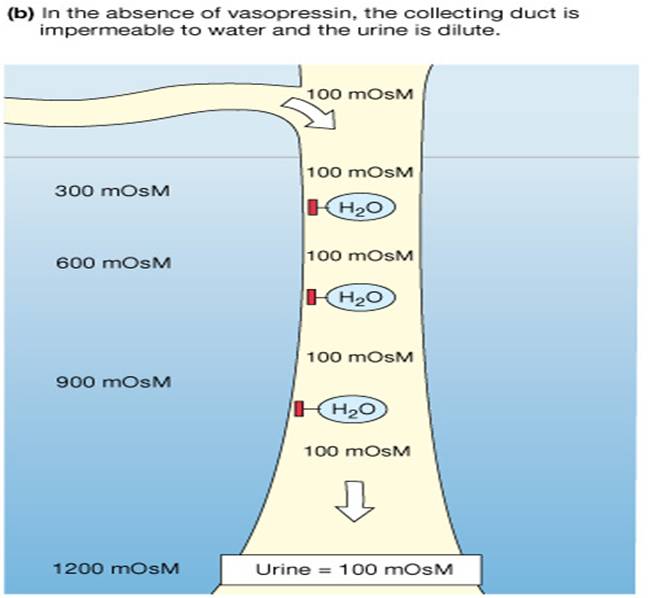
Formation of Dilute Urine
- Filtrate is diluted in the ascending loop of Henle if the antidiuretic hormone (ADH) or vasopressin is not secreted
- Dilute urine is created by allowing this filtrate to continue into the renal pelvis
- Collecting ducts remain impermeable to water; no further water reabsorption occurs
- Sodium and selected ions can be removed by active and passive mechanisms
- Urine osmolality can be as low as 50 mOsm (one-sixth that of plasma)
|
 |
Formation of Concentrated Urine
- Antidiuretic hormone (ADH) inhibits diuresis
- This equalizes the osmolality of the filtrate and the interstitial fluid
- In the presence of ADH, 99% of the water in filtrate is reabsorbed
- ADH-dependent water reabsorption is called facultative water reabsorption
- ADH is the signal to produce concentrated urine
- The kidneys’ ability to respond depends upon the high medullary osmotic gradient
|
 |
Renal Clearance
- The volume of plasma that is cleared of a particular substance in a given time:
RC = UV/P
RC = renal clearance rate
U = concentration (mg/ml) of the substance in urine
V = flow rate of urine formation (ml/min)
P = concentration of the same substance in plasma
- Renal clearance tests are used to:
Determine the GFR
GFR= concentration in urine X volume of urine per unit of time
Plasma concentration
- Detect glomerular damage
- Follow the progress of diagnosed renal disease
Creatinine Clearance
- Creatinine clearance is the amount of creatine in the urine, divided by the concentration in the blood plasma, over time.
- Glomerular filtration rate can be calculated by measuring any chemical that has a steady level in the blood, and is filtered but neither actively absorbed or excreted by the kidneys.
- Creatinine is used because it fulfills these requirements (though not perfectly), and it is produced naturally by the body.
- The result of this test is an important gauge used in assessing excretory function of the kidneys. For example grading of chronic renal insufficiency and dosage of drugs that are primarily excreted via urine are based on GFR
- Other methods involve constant infusions of inulin or another compound, to maintain a steady state in the blood.
Physical Characteristics of Urine
- Color and Transparency
- Clear, pale to deep yellow (due to urochrome)
- Concentrated urine has a deeper yellow color
- Drugs, vitamin supplements, and diet can change the color of urine
- Cloudy urine may indicate infection of the urinary tract
- pH
- Slightly acidic (pH 6) with a range of 4.5 to 8.0
- Diet can alter pH
- Specific Gravity
- Ranges from 1.001 to 1.035
- Is dependent on solute concentration
Chemical Composition of Urine
- Urine is 95% water and 5% solutes
- Nitrogenous wastes include urea, uric acid, and creatinine
- Other normal solutes include:
- Sodium, potassium, phosphate, and sulfate ions
- Calcium, magnesium, and bicarbonate ions
- Abnormally high concentrations of any urinary constituents may indicate pathology
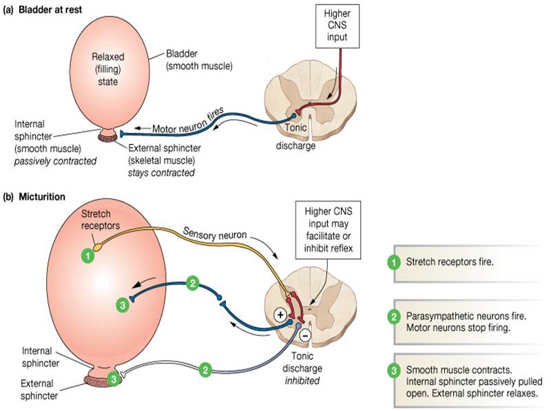
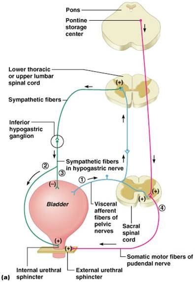
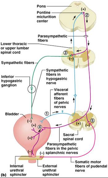
Micturition
|