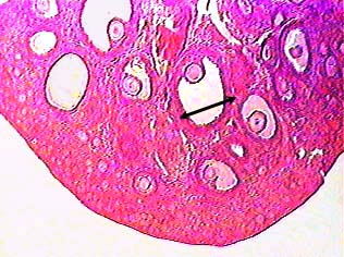
The image shows part of a section of the ovary. Follicles consisting of an oocyte surrounded by layers of follicle cells develop in the cortex of the ovary. The arrow bar shows the diameter of one mature or Graafian follicle. Many smaller follicles are in the space between the mature follicles and the outer edge of the ovary, but they just look like pink spots in this image.
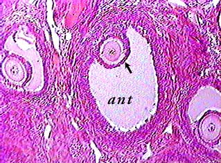
Mature human follicles are almost an inch (about 2.5 cm) in diameter. They are characterized by a fluid-filled cavity called the antrum (ant) which first appears in the secondary follicle stage. In a more mature follicle, the oocyte would look as if it were sitting on a stalk of follicle cells. The layer of follicle cells that surrounds the oocyte is called the corona radiata (arrow).
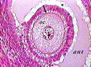
A thick layer of glycoproteins called the zona pellucida (arrow) surrounds the oocyte (oc) and separates it from the follicle cells in the corona radiata. The white bumpy areas (*) between the vesicular fluid in the antrum and the outer part of the follicle are an artifact.
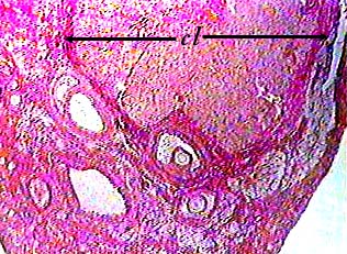
Following ovulation, the cells that remain in the follicle undergo a transformation into lutein cells. The transformation includes an increase in size and a conversion of the cytoplasm to secrete large amounts of steroids (progesterone and estrogens). The corpus luteum (cl) is easy to find because it is very large--usually larger than a mature follicle--and you can usually see some developing follicles around it.