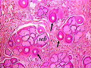
The images on this page are from a cross section of the scalp. The tissue was cut parallel to the surface of the skin. It shows several clumps of hair follicles (arrows) and sebaceous glands (seb).
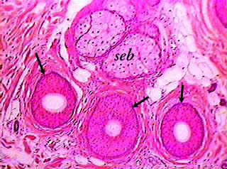
The dark circular structures with the light centers are cross sections of hair follicles containing hairs. The hair is the light area in the center of each follicle. The nuclei of the follicle cells look like darker dots.
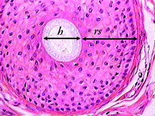
There is probably a diagram in your textbook showing a cross
section of a hair and follicle. You should compare the diagram
to this image.
One bar indicates the width of the hair (h) and the other
the epidermal root sheath (rs). The outer layer of the epidermal
root sheath is easy to recognize because the cells are cuboidal
epithelial cells and their nuclei are lined up. This layer corresponds
to the stratum basale of the epidermis. Outside of the epidermal
root sheath is a connective tissue root sheath.