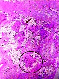
In this image you are looking at the dermis (top part of image)
and hypodermis (bottom part of image) of the skin.
Sudoriferous or sweat glands are coiled tubular glands. When
you see one on a microscope slide, it will look like a clump
of circles. The circle on this image shows the location of an
eccrine sudoriferous gland. The duct of the gland is a tube that
goes from the gland to the surface of the skin (which is not
visible on this image--it would be above the tissue shown here).
It winds back and forth as it goes towards the surface, so you
usually only see it as a series of circular structures between
the gland and the surface of the skin. The arrow in this image
is pointing to part of the duct of a sudoriferous gland.
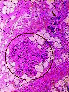
In the circle you should be able to several cross sections of the sweat gland. The light spaces are adipose cells. The best way to locate the sweat gland cells is to look for their nuclei, which are the dark dots. Each group of nuclei indicates one section of the tubular gland. The arrow is pointing to an oblique section of the cut of this gland. You can see other sections of the duct between the arrow and the circle.