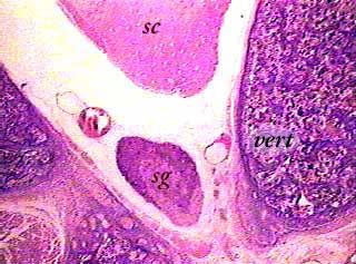
This image shows part of a cross section of the spinal column and spinal cord (sc). You can see the body of the vertebra (vert) on the right, and part of the arch on the left (the arch is not labeled). As you can tell from the location of the structures, this image does not show the spinal cord in anatomical position--the image is turned on its side. A spinal ganglion (sg) is the same thing as the dorsal root ganglion that is described in your textbook. There is one pair of ganglia (one left, one right) for each spinal nerve. They are located in an enlarged region of the dorsal spinal root and contain the cell bodies of sensory neurons that enter the spinal cord through the dorsal root.
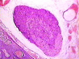
The spinal ganglion is now in the center of the image. The nuclei of some of the sensory neuron cell bodies are visible, but you may have a hard time telling them apart from artifacts caused by digitizing the image. In general, the areas of the ganglion that are darker in this image (especially upper left) are where the sensory cells are located. The areas that are lighter (lower right) are where the processes of the sensory cells are located.
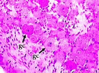
The arrows point to sensory neuron cell bodies, also called called ganglion cells (gc) in the dorsal root ganglion, or spinal ganglion. The lower left corner of the image shows and area that contains mainly nerve cell processes. Each sensory nerve cell body is surrounded by supporting cells called satellite cells. They are difficult to see in this image, but their nuclei can be seen around the outside edges of the ganglion cells.