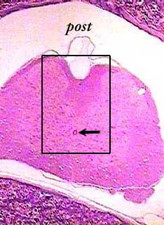
Cross sections of the spinal cord are so large that you will not be able to see the whole thing on the microscope--you will have to move back and forth or use a dissecting microscope. This section is from a slide that includes both the spinal cord and a vertebra. You are looking at the spinal cord in anatomical position, with the anterior or ventral part at the bottom and the posterior (post) or dorsal part at the top. The arrow points to the central canal, which is a good landmark to look for if you get lost when looking at this slide on the microscope. You can see the gray matter (lighter color) in the center of the spinal cord and the white matter (darker color) around the outside.
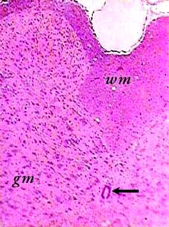
This image is an enlargement of the area inside the box in the diagram above. The central canal (see arrow) is still visible near the bottom of the image. The difference between gray (gm) and white matter (wm) is more obvious here. Most of the dark spots are the nuclei of neurons in the gray matter and supporting cells in both gray and white matter.
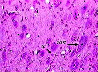
The largest cells in the spinal cord are motor neurons (mn). The small dark spot inside the cell is its nucleus. These cells are located mostly in the anterior or ventral gray matter. Because this is a section, and not a smear, you cannot see the processes. The small dark spots are the nuclei of supporting cells (sc).