
The cochlea is a coiled structure, so in this image you are looking at cross sections of several different turns. Because of the way the tissue was cut, some of the turns have very strange shapes and do not look anything like the diagrams in your text books. The most normal-looking section is enclosed in a box, which is enlarged in the image below. The circle encloses the basilar membrane and the organ of Corti.
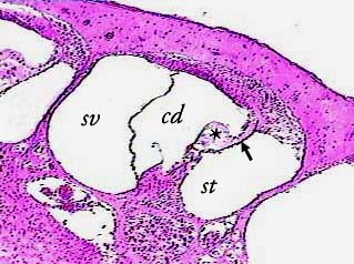
The cochlea contains a fluid-filled membrane tube--the cochlear
duct (cd)--sandwiched between two other fluid-filled canals--the
scala vestibuli (sv) and the scala tympani (st). You can see
part of the scala tympani of another "turn" of the
cochlea on the left edge of the image, and part of the scala
vestibuli of a different "turn" in the lower right
corner.
The cochlear duct and scala tympani are separated by the basilar
membrane (arrow) . Inside the cochlear duct and sitting on the
basilar membrane is the organ of Corti (asterisk).
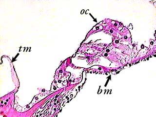
The basilar membrane (bm) forms a sort of floor for the organ of Corti (oc) to sit on. The tectorial membrane (tm) in this image it not in its normal position--it should be sitting on top of the organ of Corti (see the diagrams in your textbook).
When the cochlea is prepared and sliced to make microscope slides, a certain amount of distortion occurs. Although these images give you a much better idea of what the cells actually look like, the locations of the small structures and the relationships between some of the structures are more accurate in the diagrams in your textbook.