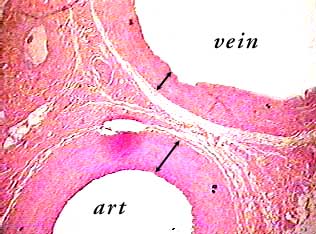
In many parts of the body, arteries and veins occur in pairs, one (the artery) taking blood to an organ and the other (the vein) returning it to the heart. In this image you can see part of an artery (art) and corresponding vein. There are several clues you can use to figure out which is which. The wall of an artery is always thicker than the wall of the corresponding vein--compare the arrow bars in the image to see the difference in wall thickness. Also the lumen of an artery is smaller than the lumen of the corresponding vein. Even though you can only see part of each vessel in the image, it is obvious that the vein has a much larger lumen.
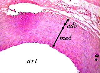
This image shows part of the artery from the image above. Its
wall has three layers: a tunica intima on the inside (see next
image for detail), a tunica media (med) in the middle, and a
tunica adventitia (adv) on the outside. The outer part of the
tunica adventitia
This is a muscular artery, which means that most of the tissue
in the tunica media is smooth muscle. All three layers contain
elastic fibers.
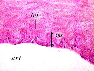
Arteries have a distinctive layer of elastic fibers called the
internal elastic lamina (iel) in the tunica intima (int). You
can see it clearly in this image as a wavy line.
Most of the dark spots you can see below the internal elastic
lamina are the nuclei of endothelial cells (the endothelium is
the layer of simple squamous epithelium that lines all blood
vessels.
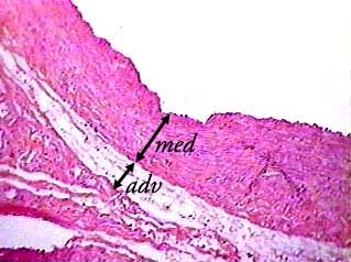
This shows part of the vein from the image at the top of the page. The tunica media (med) consists mainly of smooth muscle. The tunica adventitia of this vein is fairly thin. In some veins the tunica adventitia can be thicker than the tunica media.
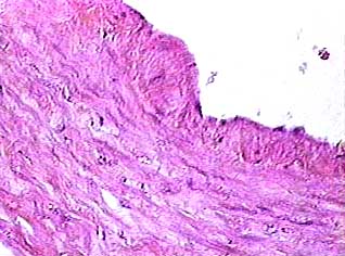
Although veins have elastic tissue in the tunica intima, it is not as obvious as the internal elastic lamina of arteries. The dark spots (arrow) on the surface are the nuclei of endothelial cells.