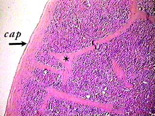
The spleen is surrounded by a tough connective tissue capsule (cap) that extends inward as trabeculae (*). Arteries, veins and nerves pass through the trabeculae.
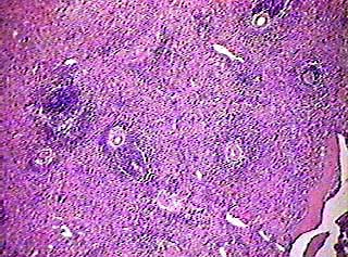
Several blood vessels can be seen in this image, as well as part of a trabecula in the lower right corner.
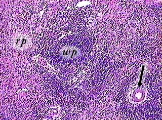
Most splenic tissue is red pulp (rp) containing red blood cells,
reticular connective tissue, macrophages and venous sinuses.
The arteries (arrow) are surrounded by white pulp (wp), which
consists of lymphocytes and reticular connective tissue. White
pulp usually looks like circular dark areas. Go back to the image
above this one and locate the white pulp.
The venous sinuses in the red pulp are usually not visible
on slides of the spleen unless the tissue is prepared by distending
it with fluid before it is fixed, embedded and sectioned.