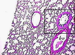
At first this might confuse you because it looks sort of like adipose tissue--they both have a lot of empty spaces. There are a lot of clues that will tell you it's lung, instead. For one thing, most of the adipose tissue you see on slides in the lab doesn't have cross sections of big tubes in it. On this image you can see two blood vessels (one labeled bv) and a bronchiole (in box), which is enlarged in the image below this one.
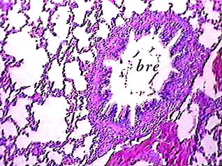
As you learned by looking at the image above this one, you will
usually see cross sections of various kinds of tubes on slides
of the lung. This is an enlargement of the bronchiole (bre) from
the image above. You have to learn to tell it apart from bronchi,
arteries and veins. (add link to artery and vein page here).
First of all, both bronchi and bronchioles have folds in the
mucosa when seen on slides (arteries and veins don't have these
folds). The folds are caused by contraction of smooth muscle
after death.
The other structures in this image are alveoli, alveolar sacs,
and alveolar ducts.
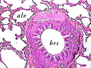
The next thing to learn is how to tell bronchi (brs) from bronchioles.
They both have the folded mucosa, so that feature won't help
in this case. But bronchi have cartilage in their walls. Here,
a plate of hyaline cartilage can be seen in the upper outer edges
of the bronchus. Relax--the image isn't good enough for you to
be able to identify the cartilage. But you should see that it
is different than the rest of the tissue forming the wall of
the bronchus.
An alveolus (alv) is also labeled in this image. Because they
are cut across at different places, it looks like there is more
variation in size than really exists.
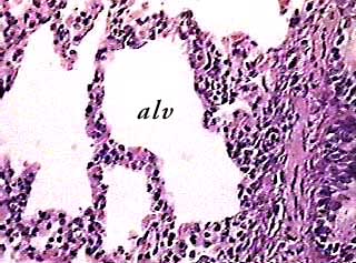
Here is a different alveolus. Now you can see the real difference between lung and adipose tissue. The outer edge of the empty space in adipose tissue consisted of the cell membrane and a very thin layer of cytoplasm. The outer edge of an alveolus shows that there are many cells in the wall of each alveolus. Most of the dark dots in this image are the nuclei of cells, and the pink areas are cytoplasm.
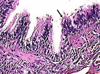
This image shows part of the wall of a bronchiole. The epithelium that forms its innermost layer is simple columnar ciliated. Although the cilia are not very clear on this image, you can see them above the surface of the cells at the arrow and around the bottom of the fold to the left of the arrow.
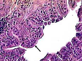
As bronchioles get smaller, the epithelium gets shorter. These cells from a smaller bronchiole than the one in the image above are simple cuboidal. That should tell you that this is a very small
bronchiole.