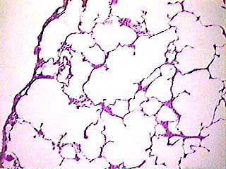
These images will look a little different from the ones on the other lung page. Because these images were made from a thin section slide, there is less tissue and more open space overall. This image does not contain any large blood vessels, bronchi or bronchioles.
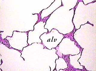
One alveolus (alv) is labeled in this image. Can you find the
other alveoli? Can you find this area on the image above?
The other spaces are probably alveolar sacs. You can see part
of the walls of the alveoli opening off of the alveolar sac.
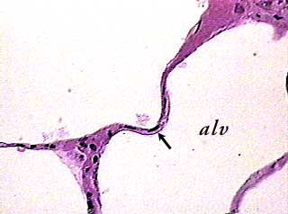
On the other lung page, the tissue was so thick that we could not see individual cells in the walls of the alveoli (alv). The arrow in this image points to the nucleus (flat dark spot) of an alveolar type I (simple squamous) cell.