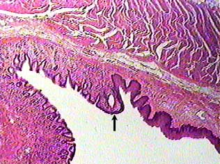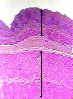
The wall of the esophagus consist of four layers: the innermost layer is the mucosa (top arrow bar in image), just outside of that is the submucosa (lighter layer between arrow bars), the next layer is the muscularis (lower arrow bar), and the outermost layer is called the adventitia.
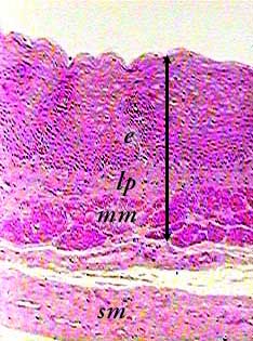
This image shows the mucosa of the esophagus (arrow bar) in more
detail. The mucosa contains three layers of tissue. The surface
of the mucosa is stratified squamous nonkeratinized epithelium
(e). Underneath or outside that is a thin layer of loose c.t.
called the lamina propria (lp). You can tell these layers apart
by looking at the density and pattern of the nuclei (dark spots)
in the two tissues. They are much closer together in the epithelium.
Below or outside of the lamina propria is the muscularis mucosae (mm), a thin layer of smooth muscle.
You can see part of the submucosa (sm) in the bottom of the
image.