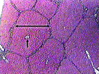
The liver is divided into hundreds of small units called lobules. In pigs, the lobules are separated from each other by connective tissue, and they are very easy to see. On this image the connective tissue looks like a dark border around the outside of each lobule. The arrow bar indicates the width of one of the lobules. Unfortunately for students of human anatomy, the human liver doesn't have these nice connective tissue partitions, so the lobules will be a lot harder to see on slides of human liver.
The arrow points to a cross section of a central vein. A central vein runs through the centers of each lobule and receives blood from the sinusoids.
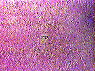
The liver can be confused with other organs that contain mainly small cells of glandular epithelium, such as the pancreas. Most of the pancreatic cells are arranged in small spherical secretory units that look like circles when sectioned. In contrast, the cells that form a liver lobule are arranged in rows that seem to radiate out from the central vein (cv). The arrangement of liver cells is not obvious in this image. Check your textbook for diagrams.
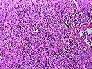
Theoretically, each liver lobule is shaped like a hexagon in cross section. At each "corner" of the hexagon there should be a branch of the hepatic artery, the hepatic portal vein and a bile duct. In reality you will not usually be able to find all of these structures at each corner of each lobule. The easiest of the three to find is the bile duct. It will be made of simple cuboidal epithelium (arrow).
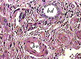
Bile ducts (bd) collect bile from the liver lobules and take it out of the liver. Where you see a small bile duct like this, you may also be able to find a small artery (bv) and a small vein.
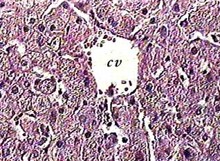
Sinusoids are special capillaries that take blood from the outside edges of the liver lobule towards the center. As blood passes through the sinusoids the liver cells can take things out of it and put other things into it. Then it drains into the central vein (cv).