
This image is a section that shows almost the entire thickness
of the kidney. The outer layer of the kidney--the cortex--is
on the top. As you move down through the image you will see part
of the kidney medulla.
The round structures in this part of the kidney are called
renal corpuscles (rc). Each renal corpuscle consists of a glomerular
capsule (part of the nephron) surrounding a clump of capillaries
called the glomerulus. The glomerulus shrinks during tissue processing,
leaving a thin white space between it and the outer wall of the
capsule. This white space makes the renal corpuscles easy to
find. They make good landmarks for identifying renal cortex,
because that is the only place they are found. The other structures
you see are sections of various parts of the nephrons: proximal
convoluted tubules, loops of Henle, and distal convoluted tubules,
and collecting tubules or collecting ducts.
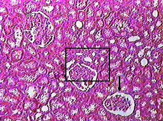
This image shows part of the kidney cortex and includes three examples of renal corpuscles. The arrow points to one of them. Most of the other things in this image are cross sections of proximal or distal convoluted tubules. The area in the box is enlarged in the image below.
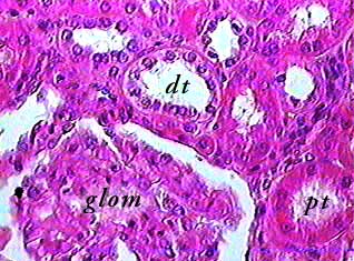
The glomerulus is easy to identify because on most slides you will see the white space of the glomerular capsule around it. Remember that this space is at least in part an artifact of tissue preparation. The proximal (pt) and distal (dt) convoluted tubules can be differentiated by looking at the cells that make up the walls of the tubules and the diameter of the lumen. Proximal convoluted tubule cells are taller, making the lumen smaller. The have long microvilli, which help to give the inside of the tube a fuzzy appearance. The distal convoluted tubule cells are shorter and do not have microvilli, so the lumen is larger.
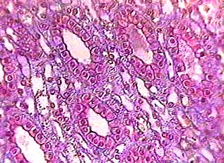
This image shows the medulla of the kidney. The specimen on this slide was cut at right angles to the one shown in the first image on this page. The large tubes made of simple cuboidal epithelium are the collecting tubules--also called collecting ducts. Between them are sections of many tubes made of simple squamous epithelium. Some of these are capillaries of the vasa recta and some are thin segments of the loop of Henle.