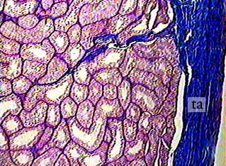
The testis is surrounded by a thick connective tissue capsule called the tunica albuginea (ta). In this image, the connective tissue components of the testis appear as dark blue because of the special stain that was used. You can see a septum formed by an inward extension of the tunica albuginea in the upper right part of the image. The circular structures are sections of seminiferous tubules.
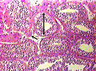
The arrow bar indicates the diameter of a seminiferous tubule.
Each tubule is surrounded by a fibrous connective tissue tunic.
In the image this layer appears as a pink circle around the outside
of the tubules. Inside the connective tissue tunic is a thin
basal lamina, and inside that is the germinal epithelium. Spermatogenesis
occurs in the seminiferous tubules.
The arrow points to a clump of interstitial cells, also called
Leydig cells. They are located outside the seminiferous tubules
and their function is secretion of testosterone.
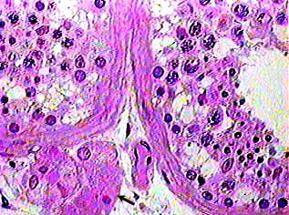
This image shows portions of two seminiferous tubules and some
interstitial cells (arrow). Within the seminiferous tubules are
two cell types: 1) supporting cells called Sertoli or sustentacular
cells, and 2) spermatogenic cells.
The sustentacular cells are almost impossible to identify because their lateral borders are extended to surround the spermatogenic cells. The only way to find one is to look for its nucleus, which should be longer and more triangular in shape than the nuclei of the spermatogenic cells. Even in a thick section, few of the sustentacular cells in a section of the seminiferous tubule would be cut through the nucleus, and so they are in effect invisible.
The spermatogenic cells form 4 to 8 layers in the walls of
the seminiferous tubules. The stem cells are called spermatogonia,
and they form the outermost layer of cell, right next to the
outer wall of the tubule. Generally their nuclei are round and
very dark. These cells undergo cell division--mitosis and then
meiosis--so you can usually detect chromosomes in the nuclei
of at least some of them in any given tubule. The nuclei of dividing
cells will look as if they contain lots of very dark specks.
The spermatogenic cells and their nuclei get smaller as they
move from the outer wall of the tubule towards the lumen.