Primate small intestine
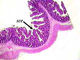
You are looking at part of the small intestine. The inner surface of the intestinal wall is made of simple columnar epithelium (sce). Instead of being smooth, the inside of the intestine is folded and covered by millions of tiny projections called villi. On the image, you can see one fold on the right and parts of several villi sticking up from the surface of the fold.
Primate small intestine
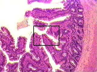
At 100X you can begin to see the layer of simple columnar epithelium. the best place to look for it is on the surface of the villi. You can see what look like round white bubbles in the epithelium. These are goblet cells and they secrete mucus. The area inside the box is the portion of this image that you will see at 400X. After you have looked at the 400X images and identified the layer of epithelium and the goblet cells, go back and look at the same things again on this image. sometimes it is easier to find something after you have seen it at a higher magnification.
Primate small intestine
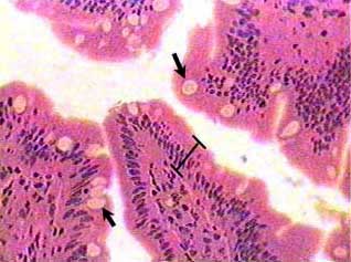
The arrows are pointing to goblet cells that produce and release mucus. They look clear because the molecules in the mucus do not absorb a lot of stain. The entire layer of simple columnar epithelium is indicated by the bar.