100X
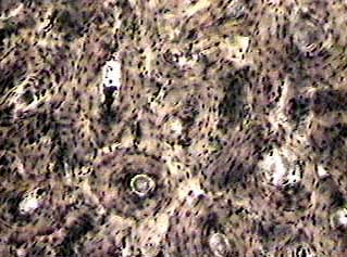
On this image you can see several of the structural units of
bone tissue (osteons or Haversian systems). Each osteon looks
like a ring with a light spot in the center. The light spot is
a canal that carries a blood vessel and a nerve fiber. The darker
ring consists of layers of bone matrix made by cells called osteoblasts
(check your textbook for an explanation of the difference between
osteoblasts and osteocytes). Between the osteons are layers of
bone matrix that don't have the circular shape. They may be the
remnants of osteons that are being remodeled into new bone tissue.
See how many osteons you can pick out in this image. There is
a good one in the lower center of the image.
Compact bone is very different from the other tissues you have seen. This image was made from a cross-section specimen of bone that has been ground to a very thin plate. Slides have to be made this way because the matrix of bone is too hard to be cut with a knife as the other tissues are. Another way of preparing bone slides is to remove the calcium salts from the matrix and then make sections by cutting off thin slices with a knife. Ground bone preparations are still very thick, and not much light can pass through them. They are excellent for showing the laminar (layered) structure of compact bone matrix, and the canals that link the osteocytes.
400X
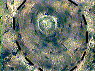
This image is from a different slide than the other two images on this page. That's why the color looks different. We have added a dotted line around the outside of the osteon in case you had trouble picking them out on the previous image. Notice the layered effect in the matrix. The layers of matrix (lamellae) are added to the outside of the osteon as it grows larger. The amount of matrix that can be added to an osteon is limited by the distance that nutrients can diffuse to the osteoblasts from the blood vessel in the central canal.
400X
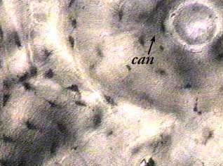
In this image, the canal is the light spot in the upper right corner. The dark spots are the lacunae where osteocytes would normally be found. The osteocytes themselves are not preserved in this type of preparation. Look at the lacunae in the lower left corner of the image. The small wavy lines that connect them are the canaliculi that connect osteocytes to each other and to the central canal in living bone tissue.