Cardiac muscle 40X
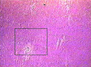
The individual cardiac muscle cells are arranged in bundles that form a
spiral pattern in the wall of the heart. On any slide of cardiac muscle
you will see cells that have been sectioned in every possible direction,
from transverse to oblique to longitudinal. The cells and their detailed
structure is best seen on cells that are sectioned longitudinally. While
you are on low power, scan the slide for an area where the cardiac muscle
cells seem to be the longest.
The area in the box is enlarged in the next image.
Cardiac muscle 100X
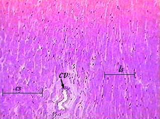
In this image, you can see cells sectioned longitudinally (ls) and transversely (cs). In the lower part of the image you can see a coronary blood vessel (cv).
Cardiac muscle 400X
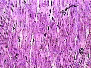
Cardiac muscle cells branch and attach to each other. The circle indicates
a place where two cardiac cells are branched and connected to each other.
Some of the nuclei (nuc) will look round and others will look flat. It all
depends on how the cell was cut.
The striations in cardiac muscle are not as obvious as those of skeletal
muscle. But you can see faint lines running across the cells in this image.
If you have trouble seeing these on your slide in lab, close the iris diaphragm
a little to increase the contrast.
Cardiac muscle 400X
showing intercalated disks
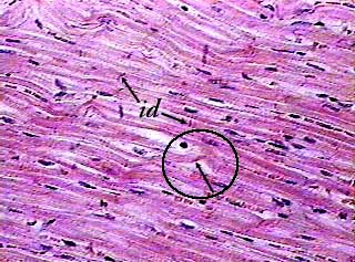
Cardiac muscle cells are joined end to end at special junctions called intercalated
discs (id). These appear as dark lines that are perpendicular to the axis
of the cell (they run across the cell). If you have trouble finding them
in lab, first increase the contrast by closing the iris diaphragm a little,
then use the fine focus knob to focus up and down. As you do this, structures
that are thinner than the tissue section will come into and go out of focus.
You should be able to see hundreds of intercalated discs on each slide.
The circle and arrow indicate another point where two cells are branched
and interconnected.