Content
Nervous System Review
Nervous tissue
Cerebrospinal Fluid (CSF)
Blood-Brain Barrier
Major Areas of the Brain
---Cerebrum
---Functional Areas of The Cortex
---Motor Cortex
---Language Areas
---Basal Ganglia
---Thalamus
---Hypothalamus
---Limbic System
---Cerebellum
---Brain Stem
---Pons
---Medulla Oblongata
Reticular Formation
ElectroEncephaloGram (EEG)
Memory
Basic Learning Behavior
Long-Term Potentiation
Memory Processing
Spinal Cord
Gray Matter
White Matter
Neural Reflexes
Reflex Arc
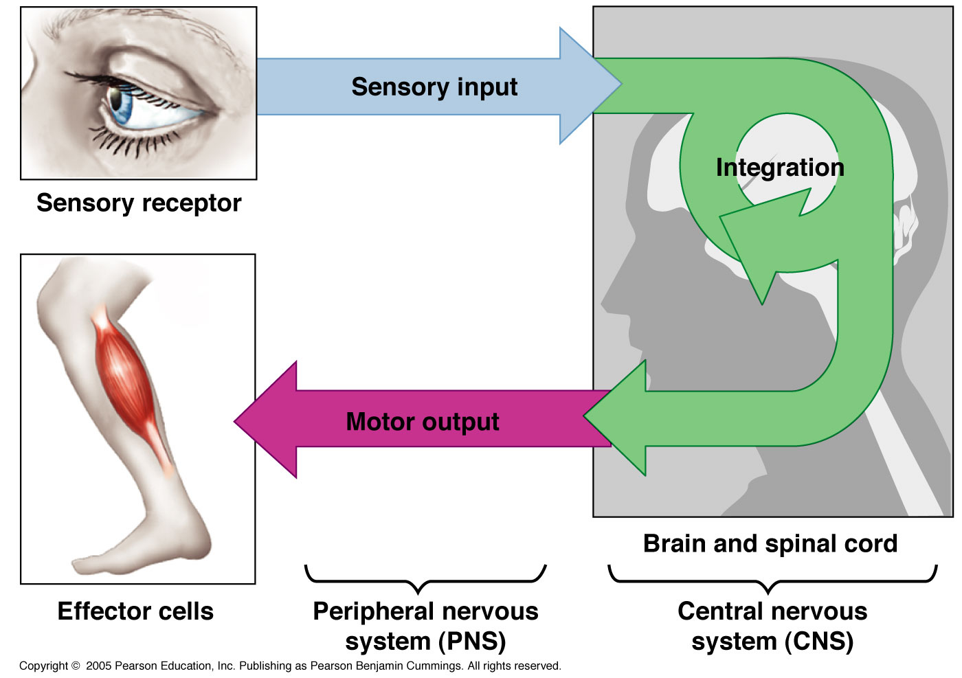
Nervous System
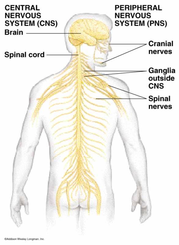
The nervous system has 2 major divisions:
1) Central nervous system (CNS) - consists of the brain and spinal cord. Functions to integrate and correlate sensory information; generates thought, perception, and emotions; forms and stores memory; regulates most of the body’s physiology and movement
2) Peripheral nervous system (PNS) - consists of the spinal nerves, cranial nerves, and ganglia. Functions to carry messages to and from the spinal cord and brain
Terminology review
Nerve fiber - a general term for any neuronal process
Nerve - a bundle of many nerve fibers along the same path in the PNS surrounded by a connective tissue layer.
Ganglia - a cluster of nerve cell bodies in the PNS
Nucleus - a mass of cell bodies and dendrites in the CNS
Tract - a bundle of nerve fibers along the same path in the CNS without a connective tissue layer.
White matter - aggregations of myelinated processes from many neurons
Gray matter - contains either neurons cell bodies, dendrites, and axon terminals, or unmyelinated axons and neuroglia
There are 3 classes of neurons
1. Afferent – transmit sensory impulses from PNS to the CNS.
- Sensory afferent fibers – carry impulses from skin, skeletal muscles, and joints
- Visceral afferent fibers – transmit impulses from visceral organs
2. Efferent - transmit motor impulses from CNS to PNS
- Somatic nervous system – provides conscious control of skeletal muscles
- Autonomic nervous system – regulates smooth muscle, cardiac muscle, and glands
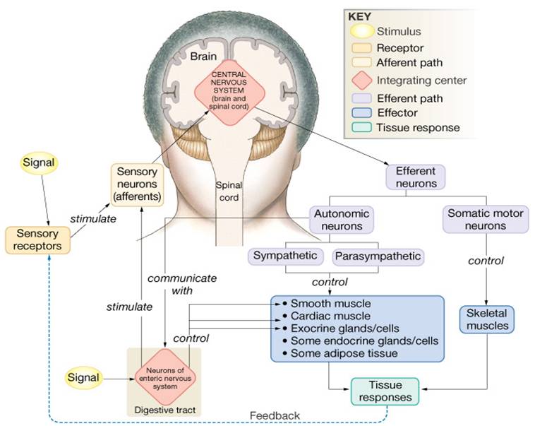
3. Association neurons or interneurons All other neurons are termed as association neurons or interneurons responsible for integrating afferent information and formulating an efferent response to include higher cognitive functions
Nervous tissue
Nervous tissue is made up of:
- Neuroglia are specialized nervous tissue cells that are smaller and more numerous than neurons. They serve to carry out support functions such as vascularization, phagocytosis and myelinization.
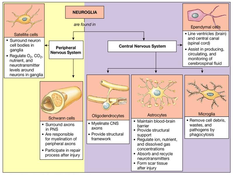
There are four types of neuroglia found in the CNS:
1. Astrocytes - star shaped with many processes, participate in the metabolism of neurotransmitters, maintain proper K+ balance, help form blood brain barrier, and provide a link between neurons and blood vessels.
2. Oligodendrocytes - fewer processes and are smaller that astrocytes, most common type in the CNS, they are involved in myelinization.
3. Microglia - small cells derived from monocytes that function as macrophages and carry out phagocytosis.
4. Ependymal - columnar to squamous epithelial cells often ciliated that form a lining in the ventricles (fluid filled cavities in the brain).
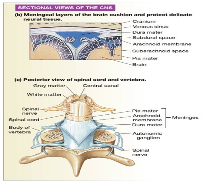
CNS Protection
- Cranial bones
- Vertebral column
- Meninges (dura mater, arachnoid, pia mater)
- Cerebrospinal fluid (CSF)
Cerebrospinal Fluid (CSF)
- CSF is a clear, colorless liquid that nourishes and protects the CNS from chemical and physical injury
- It circulates through the subarachnoid space around the brain and spinal cord and through the four ventricles (cavities) within the brain
- It contains glucose, proteins, lactic acid urea, ions, some lymphocytes.
- - Formed by selective transport across ependymal cells
- Volume 125-150 ml and is replaced > 3 times/day, flow maintained by 10 mmHg pressure gradient
- Path: ventricles ® subarachnoid space, reabsorbed into blood in dural sinuses through arachnoid villi
Functions of CSF:
- Shock-absorbing medium
- Provides a optimum and stable environment for generating nerve impulses
- Provides a medium for the exchange of nutrients and wastes between blood and nervous tissue.
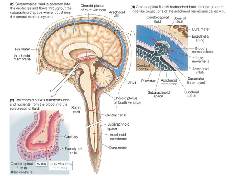
Production of CSF
- The choroid plexus is a network of blood capillaries in the walls of the ventricles
- The capillaries are covered by ependymal cells that produces the CSF.
- The ependymal cells provide a fluid tight barrier around the capillaries called the blood-brain barrie
Click here for an animation that describes the production and flow of cerebrospinal fluid.
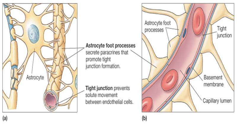
Blood-Brain Barrier
- Formed by capillary endothelial cells formed with tight junctions
- The tight junctions restrict transport between cells
- Astrocytes induce formation of tight junctions and help control transcellular transport such as K+
- Some materials pass the blood-brain barrier
- - Materials that diffuse through the lipid bilayer cannot be restricted –O2, CO2, alcohol, steroids, H2O
- Materials that require carrier transport are restricted – glucose, amino acids, and ions
- Some materials cannot pass the barrier – potentially harmful substances, some hormones/drugs
- Functions of the blood-brain barrier:
- - Protects CNS from chemical fluctuations
- Prevents entry of harmful substances
- Prevents entry of molecules that could act as neurotransmitters
- There are only 3 places in the brain that do not have a blood brain barrier:
1. Choroid plexuses (because they make CSF)
2. Hypothalamus (needs to let hormones into bloodstream)
3. Pineal gland (also needs to let hormones into the bloodstream)
Major Areas of the Brain
- About one trillion interneurons fill the brain
- Each interneuron has up to 200,000 synapses each
- There are several major areas of the brain:
- Cerebrum (including the cerebral cortex and basal nuclei)
- Thalamus
- Hypothalamus
- Cerebellum
- Brain Stem
Click here for an animation that summarizes the major areas of the brain and the function of each area.
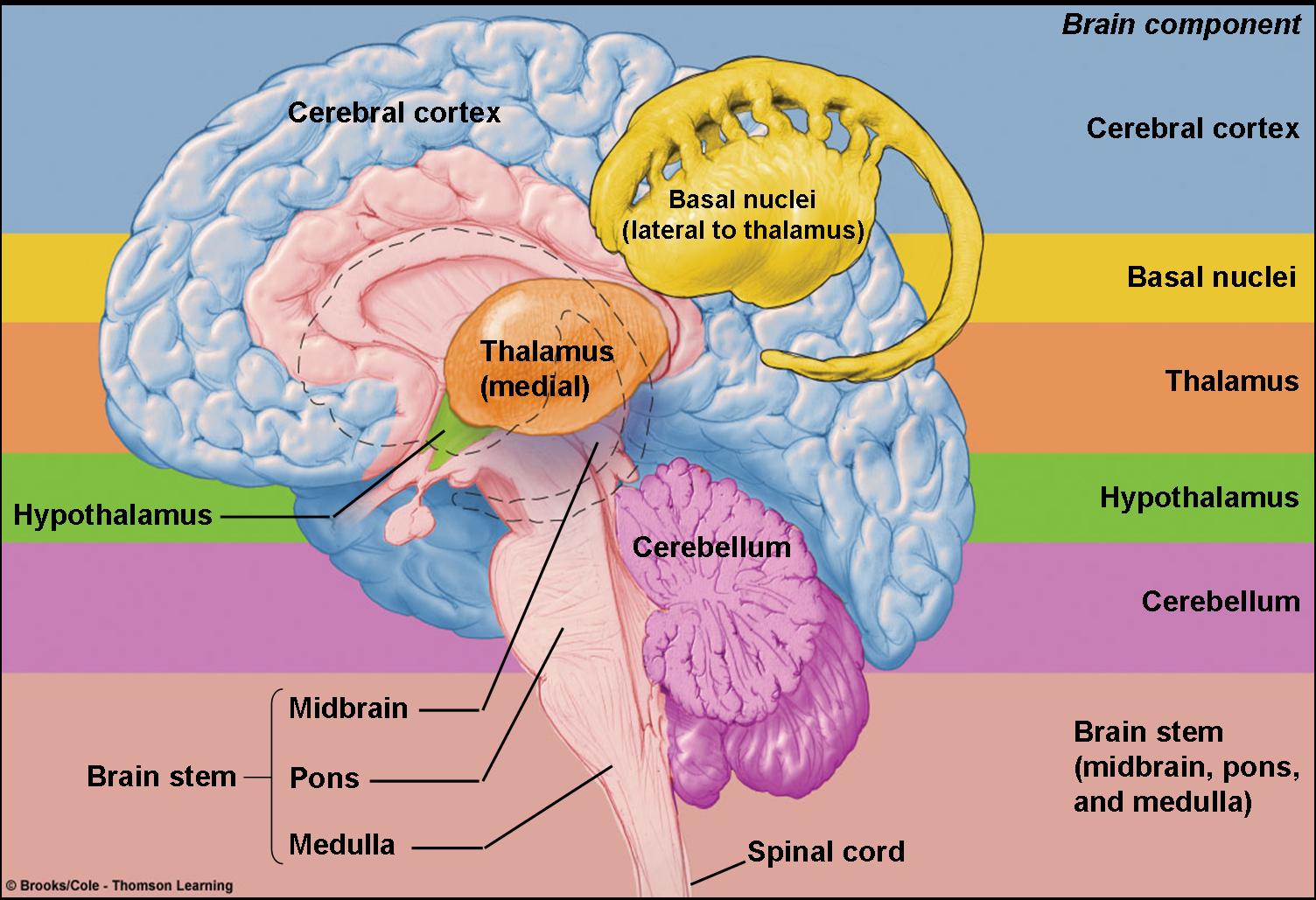
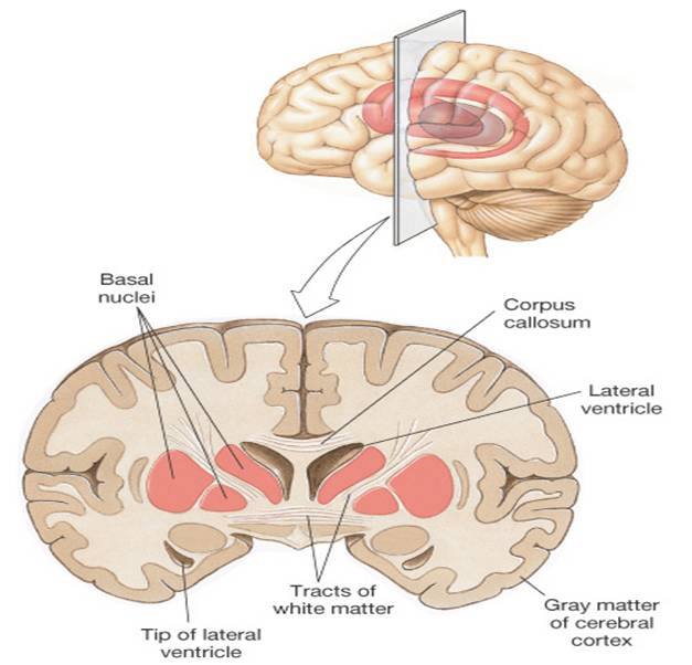
Cerebrum
- Includes cerebral cortex, tracts, and basal nuclei
- Is highly developed
- Makes up about 80% of total brain weight (largest portion of brain)
- The inner core houses basal nuclei
- The o uter surface is made up of the highly convoluted cerebral cortex:
- Highest, most complex integrating area of the brain
- Plays key role in most sophisticated neural functions
Cerebral Cortex - (gray matter “computer”)
- Each half of the cerebral cortex is divided into four major pairs of lobes
- Occipital Lobes - carries out initial processing of visual input
- Temporal Lobes - initial reception of sound sensation
- Parietal Lobes - somatosensory processing
- Frontal Lobes responsible for
- Voluntary motor activity
- Speaking ability
- Elaboration of thought
- The outer layer of cerebrum hold the soma or cell bodies of the neurons.
- Surface features of the cerebral cortex include gyri (ridges), sulci (grooves), and fissures (deep grooves)
- The fissures increase the surface area and divide the brain into the four major pairs of lobes
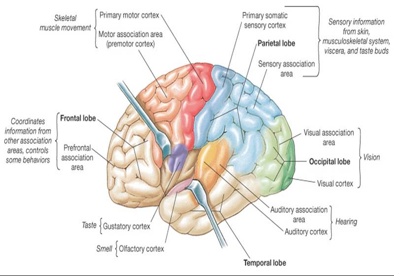
Functional Areas of The Cortex
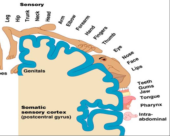
Primary Somatosensory Cortex
- Located in the post central gyrus (directly posterior to central sulcus) of each parietal lobe
- Receives input from somatic sensory receptors for proprioception, touch, pain, temperature.
- Primary function is to localize exact sites where sensations originate
- Sensory homunculus – shows proportional distribution of sensory input to the somatosensory cortex from different parts of the body based on degree of sensory perception
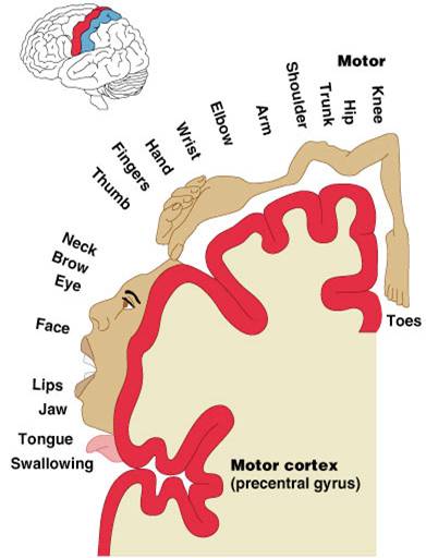
Primary Motor Cortex
- Located in the precentral gyrus of the frontal lobe
- Controls voluntary contractions of specific muscles or groups of muscles.
- Size of area and number of neurons representing each part of the body is proportional to precision and complexity of movement of that part
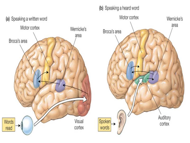
Language Areas
Comprehension and translating thought into speech involves both sensory, association, and motor speech areas located in the frontal lobe.
- Broca’s area – speech formation, speaking ability
- Wernicke’s area – language comprehension
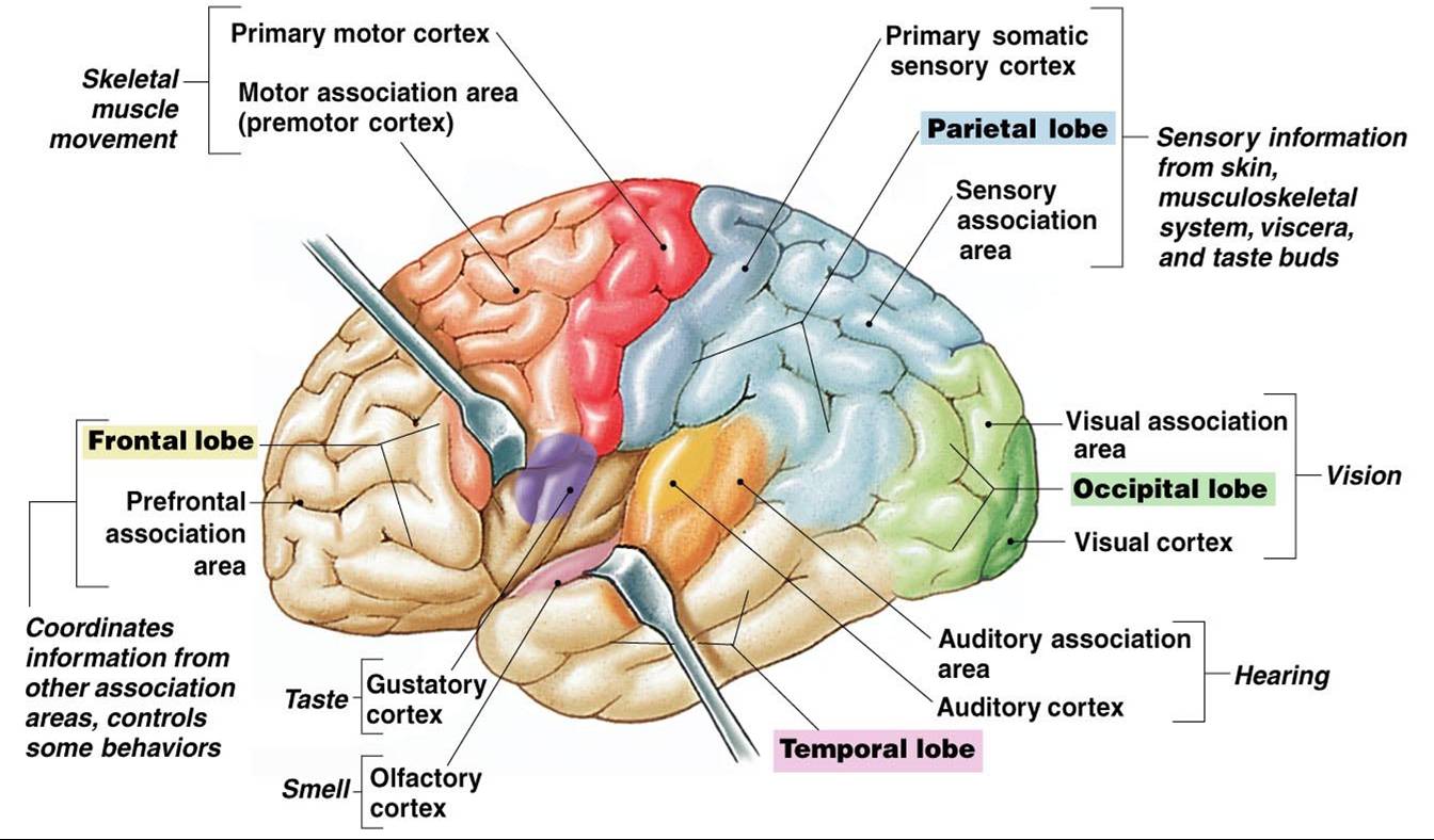
Primary Visual Cortex
- Medial surface of occipital lobe
- Receives input from the thalamus (lateral geniculate nuclei) concerning shape, color, and movement
Primary Auditory Cortex
- Is located in superior part of the temporal lobe
- Interprets characteristics of sound, hearing
Primary Gustatory Area
- Is located at the base of post central gyrus in the parietal lobe
- Receives impulses for taste
Primary Olfactory Area
- Located in the medial aspect of temporal lobe
- Receives impulses for smell
Supplemental Motor Area
- Plays preparatory role in programming complex sequences of movement
Premotor Cortex
- Orients the body and arms toward a specific target
- Deals with learned motor activities of a complex and sequential nature.
Association Areas
- Are tracts that connect motor and sensory areas and large parts of the cortex
Prefrontal Association Cortex
- Involved in planning voluntary activity, decision making, creativity, personality traits
Parietal-Temporal-Occipital Association Cortex
- Pools and integrates somatic, auditory, visual sensations for complex perceptual processing
- Also involved with language
Limbic Association Cortex
- Involved in motivation and emotion involved with memory
Integration of Cortical Regions
Sensory input >> somatosensory cortex >> higher sensory areas >> association areas >> higher motor area >> primary motor cortex >> motor output
Basal Ganglia
- Is a group of nuclei (clusters of cell bodies) in each cerebral hemisphere
- Regulates (inhibits) muscle tone required for smooth body movements
- Selects and maintains purposeful motor activity
- Monitors and coordinates slow sustained contractions – posture/support
Thalamus
- Is an oval structure consisting of gray matter organized into nuclei
- Is the principle relay station for all sensory input to the cerebral cortex from spinal cord, brain stem, cerebellum and other parts of the cerebrum.
- Routes important sensory impulses to appropriate areas of the somatosensory cortex.
- It directs attention to stimulus of interest:
medial geniculate nuclei - hearing
lateral geniculate nuclei - vision
ventral posterior nuclei - taste and somatic sensations (pain, pressure)
anterior nucleus - emotions and memory

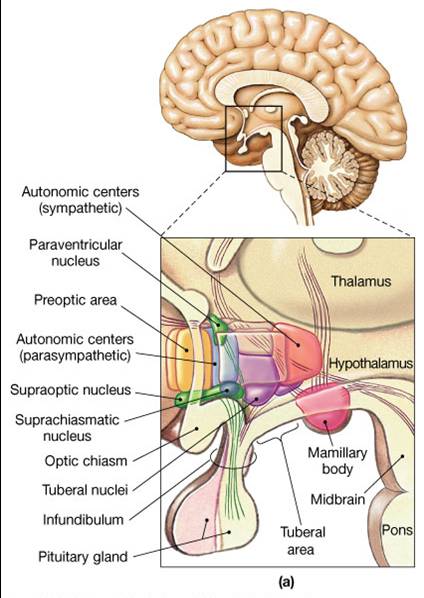
Hypothalamus
- Is located below thalamus
- Is a major regulator of homeostasis
- Controls and integrates activities of the autonomic nervous system (ANS)-regulates the contraction of smooth muscle, cardiac muscle and secretions of many glands.
- The main regulator of visceral activities such as heart rate, GI tract movement, and bladder contraction
- Associated with feelings of rage and aggression
- Regulates body temperature, food intake, and thirst
- Maintains waking state and sleep patterns
- Controls anterior pituitary hormone secretion
- Produces and transports hormones (antidiuretic hormone-ADH) to the posterior pituitary gland.
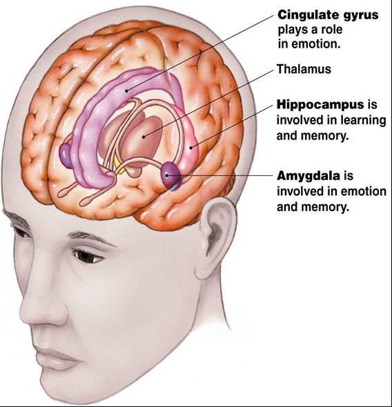
Limbic System
- Is a ring of structures encircling the brain stem
- It includes portions of the cortical lobes, amygdala, basal nuclei, nuclei of the hypothalamus and of the thalamus, and olfactory bulbs.
- Functions in the emotional aspect of behavior and in memory in conjunction with the hippocampus and cerebrum
Cerebellum
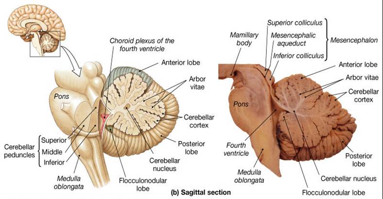
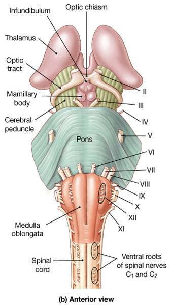
Brain Stem
- Consists of medulla, pons, midbrain
- Is an important link between spinal cord and higher brain levels relays motor and sensory impulses between other “higher” parts of the brain and spinal cord
- -Midbrain – eye movement control
- Pons/Medulla
- Signal relay
- Involuntary functions
- Many cranial nerves enter
- Pyramids – nerve tracts crossover
Pons
- Sensory and motor nuclei for four cranial nerves
- Nuclei that help control respiration
- Nuclei and tracts linking the cerebellum with the brain stem, cerebrum and spinal cord
- Ascending, descending and transverse tracts
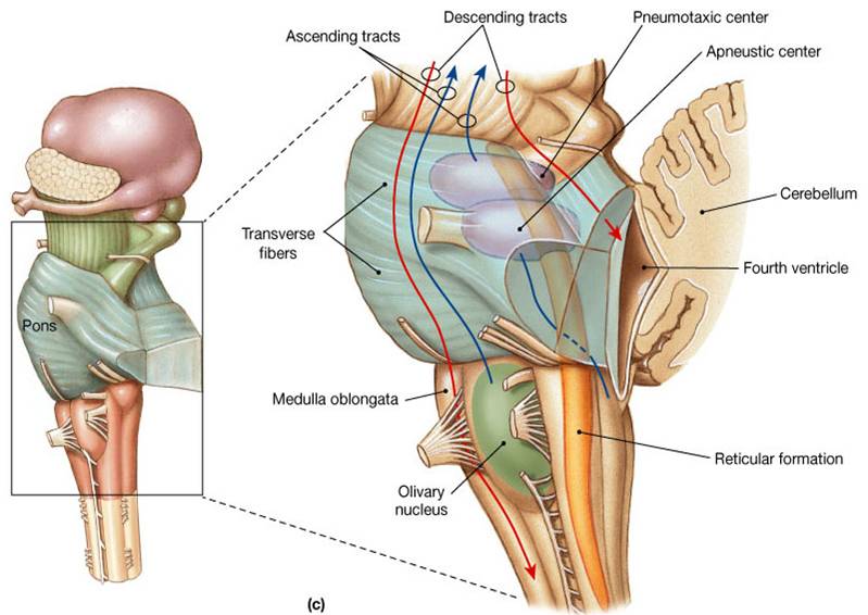
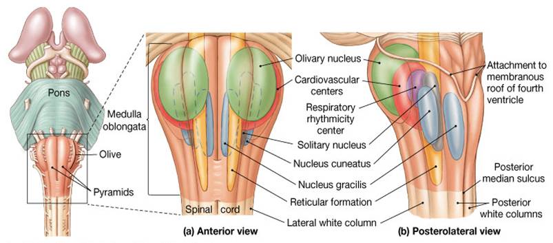
Medulla Oblongata
- Contains relay stations and reflex centers:
- -Olivary nuclei
- - Cardiovascular and respiratory rhythmicity centers
- - Cardiovascular center - regulates rate and force of heartbeat and vasoconstriction/dilation
- - Respiratory center - regulates basic breathing rhythm
- Reticular formation begins in the medulla oblongata and extends into more superior portions of the brainstem
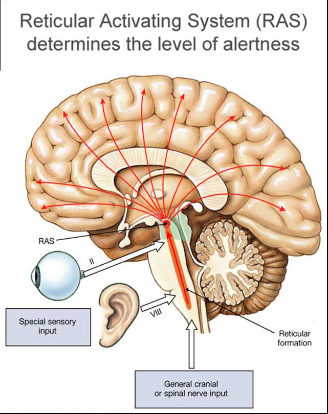
Reticular Formation
- Network of interconnected neurons runs throughout brain stem and into thalamus
- Functions in consciousness and arousal
- Receives and integrates all incoming sensory synaptic input
- Reticular activating system (RAS) - ascending fiber sends signals upward to arouse and activate the cerebral cortex, controls overall degree of cortical alertness or level of consciousness - maximum alertness >> wakefulness>> sleep >> coma
- Level of consciousness depends on the cyclical interplay between the RAS, a slow wave sleep center and a paradoxical sleep center both in the hypothalamus
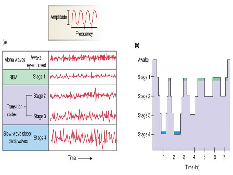
ElectroEncephaloGram (EEG)
- Records electrical activity within cerebral cortex from EPSPs and IPSPs.
- Used to diagnose:
- Cerebral dysfunction
- Brain death
- Sleep patterns
Function of Sleep
- Is a time to restore biochemical and physiological processes degraded during sleep
- Role of adenosine on sleep:
- Adenosine backbone of ATP, body’s energy molecule
- Increased levels generated during wakefulness
- Adenosine as a neuromodulator has been shown to inactivate arousal center
- Caffeine blocks adenosine receptors in the brain – prevention inhibitory action on the arousal center
- Sleep necessary to accomplish long term structural and chemical adjustments required for learning and memory, especially consolidation of procedural memories
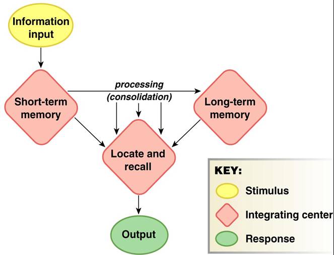
Memory
- Is "storage of acquired knowledge for later recall"
- Neural change responsible for retention and storage of knowledge is known as the memory trace
- Consolidation - Process of transferring and fixing short-term memory traces into long-term memory stores
- Working memory - Temporarily holds and interrelates various pieces of information relevant to a current mental task
- Short term memory
- Characteristics:
- Seconds to hours
- Limited capacity
- Rapid retrieval
- Mechanisms:
- Temporary modifications of synaptic function
- Changes in ion channels in the axon terminals
- Presynaptic facilitation (cAMP)
Basic Learning Behavior
Habituation – decreased responsiveness to stimulus, closure of Ca channels leads to reduced neurotransmitter release
Sensitization – increase responsiveness, release of serotonin from interneuron> increase cAMP in presynaptic neuron which blocks K channels > prolongs AP so Ca channels are open longer, increasing neurotransmitter output
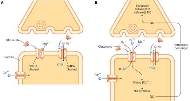
Long-Term Potentiation (LTP)
Starts with release of glutamate from activated presynaptic neuron
> AMPA receptors > EPSPs
> NMDA receptors > open Ca channels > second messenger formation > increases # of AMPA receptors
or release of nitric oxide (NO) > acts on presynaptic neuron to increase neurotransmitter release
Long Term Memory
- Last days to years
- Unlimited capacity
- Mechanisms - permanent structural changes in the brain
- Formation of new synapses between existing neurons
- Increased dendritic surface area
- Increase in neurotransmitter receptors
- Changes in neurotransmitter synthesis
- Consolidation is affected by:
- Amount of rehearsal
- Association of new & old data
- Level of excitement/importance of information
- CREB – regulatory proteins that activate genes important in long term memory storage

Memory Processing
Hippocampus and medial temporal lobe - short term memory and consolidation declarative memory (facts), specific objects, requires conscious recall
Cerebellum – “how to” memories, motor skills(procedural), subconscious recall
Prefrontal cortex - working memory, processes new and retrieved information, temporary storage, problem solving, planning, organizing
Spinal Cord
- The spinal cord is a continuation of the brain (medulla).
- Is a long cylindrical cable of nervous tissue,
- Is 1-2 centimeters in diameter and extends from the foramen magnum of the skull to L1 vertebrae.
It is about 18" (42-45 cm) long
- Is located in the vertebral canal and protected by the vertebrae
- Is surrounded by three protective connective tissue coverings - spinal meninges
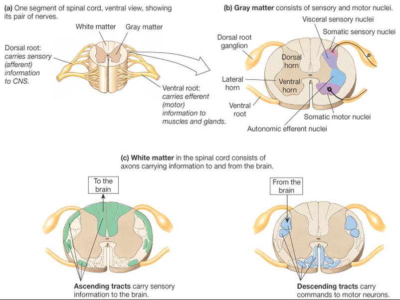
- The spinal cord, like the brain is made up of gray matter and white matter (the gray matter is centrally located in a butterfly shape)
- 31 pairs of spinal nerves originate from the spinal cord (spinal nerves are part of the PNS)
- The spinal cord serves as the major pathway for impulses to and from the brain-
- it conveys sensory impulses from the body (from sensory receptors) to the brain
- it conveys motor impulses to the body (to muscles and glands).
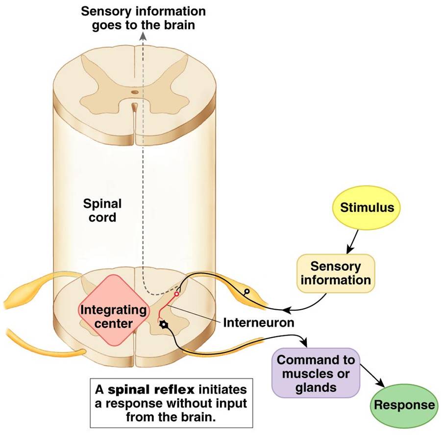
- The spinal cord also integrates information on its own, controlling spinal reflexes that occur without any brain involvement (ex: withdrawal from pain)
Gray Matter
Mostly cell bodies
- Dendrites & terminals
- Spinal reflex integrating center
White Matter
- Bundles of myelinated axons
- Ascending tracts – sensory
- Descending tracts – motor
- Dorsal roots
- Ventral roots
Extending from the spinal cord are dorsal and ventral roots which converge to form 31 pairs of spinal nerves.
Spinal nerves provide the pathway between the spinal cord and most of the body.
The core - gray matter - primarily consists of cell bodies of motor neurons and nuclei (clusters of nerve cell bodies in CNS) neuroglia, unmyelinated axons and dendrites of association and motor neurons
The gray matter is shaped like an H or butterfly formed by right and left ventral (anterior), lateral and dorsal (posterior) horns connected by the gray commissure.
The outer portion - white matter - contains myelinated motor and sensory nerve fibers to and from the brain and other levels of the spinal cord. The white matter is made up of three broad columns ( funiculi); the anterior, lateral and posterior white columns. Each column is made up of ascending (sensory) and descending (motor) tracts.
Sensory and motor tracts of the white matter
Spinal nerves are the paths of communication between the spinal cord and most of the body. Each spinal nerve attaches to a segment of the spinal cord to form roots.
Dorsal root - contains sensory nerve fibers and conducts nerve impulses from the periphery into the spinal cord.
Each posterior root has a swelling called the dorsal root ganglion, which contains the cell bodies of the sensory neurons.
Ventral root - contains motor neuron axons and conducts impulses from the spinal cord to the periphery.
Neural Reflexes
- Stimulus
- Sensory receptor
- Sensory (afferent) neuron
- CNS integration
- Efferent (motor) neuron
- Effector (target tissue)
- Response (movement)
- Feedback to CNS
Reflexes
- Are fast predictable, automatic responses to changes in the environment that help maintain homeostasis,
- Reflexes are of three types: cranial reflexes, somatic reflexes and visceral (autonomic) reflexes.
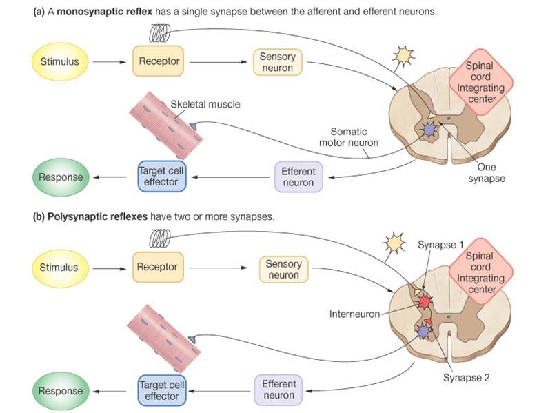
Reflex Arc -
Is the simplest pathway, involving five components
1. Receptor - distal end of a sensory neuron (dendrite) that responds to a stimulus
2. Sensory neuron/afferent pathway - conducts impulse from receptor to axon terminal located in the gray matter.
3. Integrating center in CNS - may be monosynaptic or polysynaptic involving other association neurons
4. Motor neuron/efferent - relays impulse from integrating center to part of the body that will respond.
5. Effector - gland or muscle that responds to motor neuron impulse.
Click here for an animation that describes reflex arcs.
Top ...... Main Page |