Muscular System
|
Note: Muscle accounts for nearly half of the body’s mass
- Muscles have the ability to change chemical energy (ATP) into mechanical energy |
Content
Types of Muscle Tissue
Histology Review
Skeletal Muscle Contraction (Sliding Filament Model)
Muscle Tension
Muscle Tone
Strength
Types of Contractions
Metabolism / Muscle Fatigue / Oxygen Debt
Muscle Fiber Types
Smooth Muscle
Smooth Muscle Contraction
Cardiac Muscle
Types of Muscle Tissue
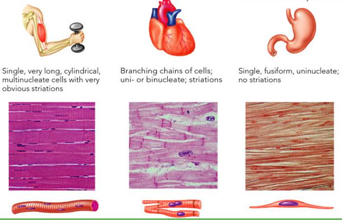
There are 3 types of muscle tissue:
- Skeletal
- Is attached to bones
- it is striated
- Is under voluntary control
- Muscle cells (fibers) are multinucleated
- Smooth
- Is located in the walls of hollow internal structures
- Is nonstriated
- Is under involuntary control
- Cardiac
- It forms most of the heart
- It is striated
- It is under involuntary control
- It is autorhythmic
Histology Review
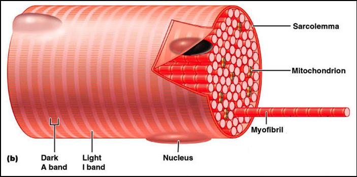
Muscle cells are called muscle fibers. Each muscle fiber is made up of:
1. Sarcolemma
- This is a muscle fiber plasma (cell) membrane
2. Sarcoplasm
- This is the muscle fiber cytoplasm
- It contains glycosomes (granules of glycogen) and the oxygen-binding protein called myoglobin
- Is is almost completely filled with contractile filaments called myofilaments
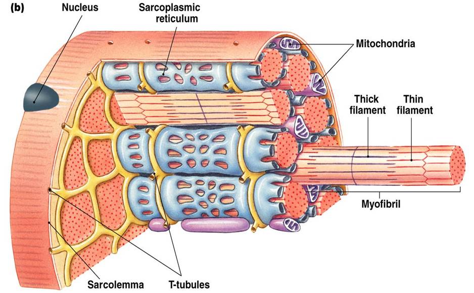
3. Sarcoplasmic Reticulum
- This is the smooth endoplasmic reticulum (SER) in the muscle fiber.
- It is a network of tubes surrounding myofibrils
- It reabsorbs calcium ions during relaxation
- It releases calcium ions to cause contraction
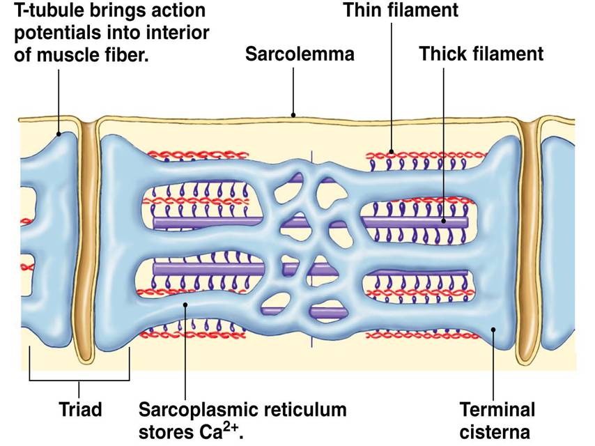
4. Transverse Tubules
- These are tubules formed by invaginations of the sarcolemma and flanked by the sarcoplasmic reticulum
- They carry action potentials deep into the muscle fiber.
- The T tubules and sarcoplasmic reticulum provide tightly linked signals for muscle contraction.
- There are T tubules at each A band/I band junction and they are continuous with the sarcolemma.
- They conduct electrical impulses to the throughout cell (every sarcomere); the electrical impulse signals for the release of Ca2+ from adjacent terminal cisternae
- T tubule proteins (Dihydropyridine) act as voltage sensors
- SR foot proteins are (ryanodine) receptors that regulate Ca2+ release from the SR cisternae
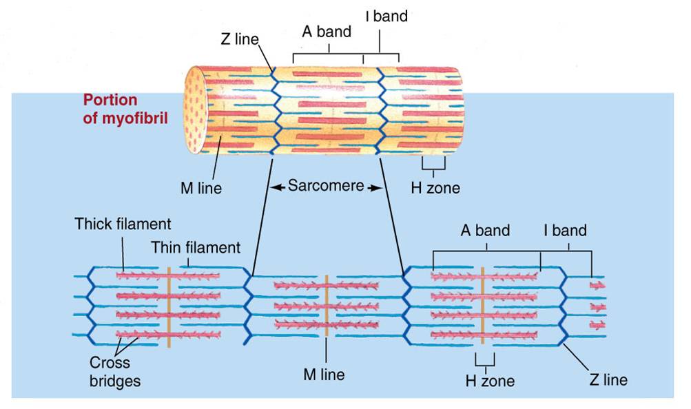
5. Myofibril
- Is a bundle of thread-like contractile elements consisting of myofilaments
- Make up 80% of the muscle volume
- Contain the contractile elements of skeletal muscle cells
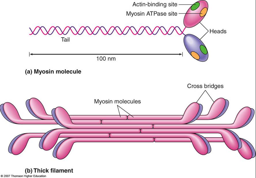
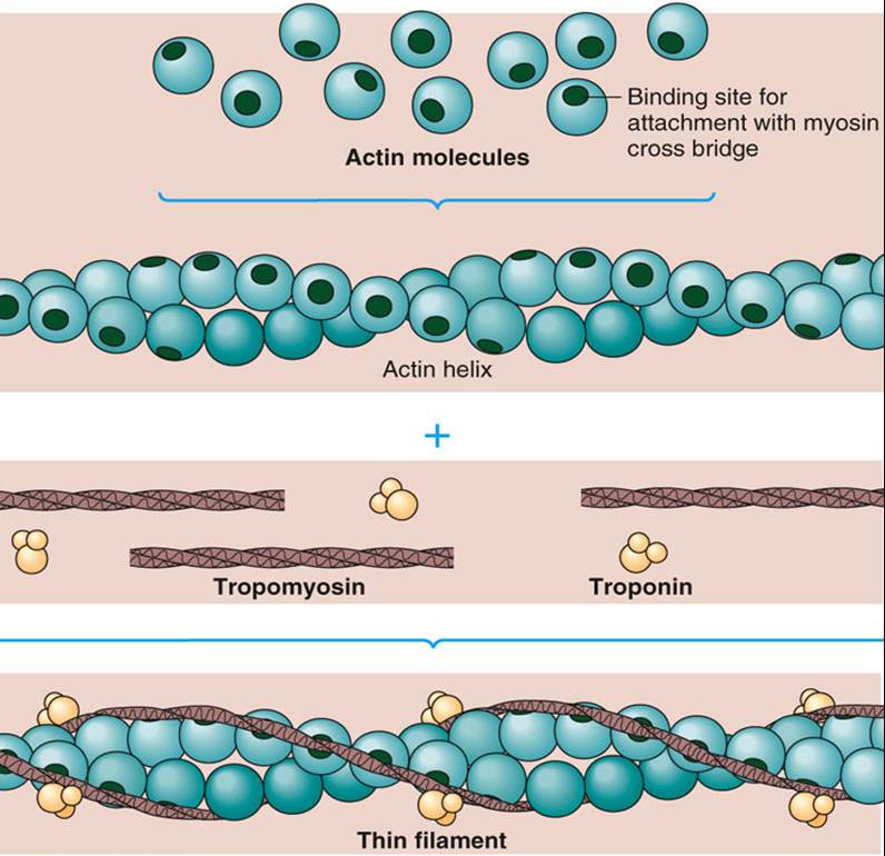
6. Myofilaments
- Are extremely fine thread like proteins
- There are 3 types of myofilaments:
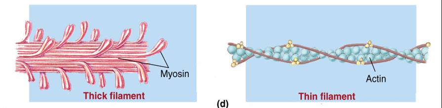
7. Sarcomere
- Is the smallest contractile unit of a muscle fiber, a compartment of myofibrils
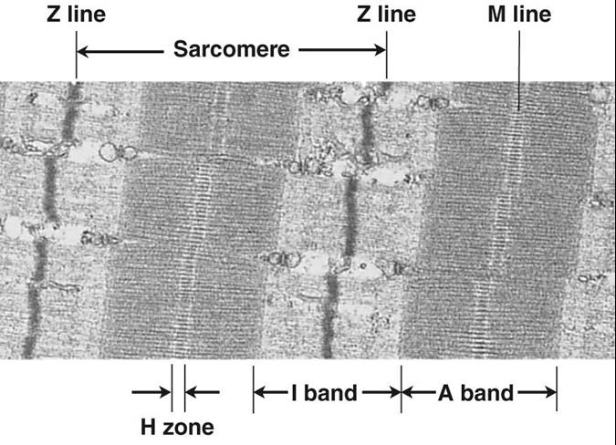
The sarcomere is characterized by alternating light and dark bands or zones produced by the myofilaments
Z disc - a line that separates one sarcomere from another
M line - central line of the sarcomere where myosin filaments are anchored
H zone - the area where only myosin filaments are present
I band - the area where only actin filaments are present
A band - includes overlapping myosin and actin filaments |
For contraction to occur, a skeletal muscle must:
- Be stimulated by a nerve ending
- Propagate an electrical current, or action potential, along its sarcolemma
- Have a rise in intracellular Ca2+ levels, the final stimulus for contraction
Click here to review what happens to each of the parts of a sarcomere during muscle contraction. The animation is followed by practice questions.
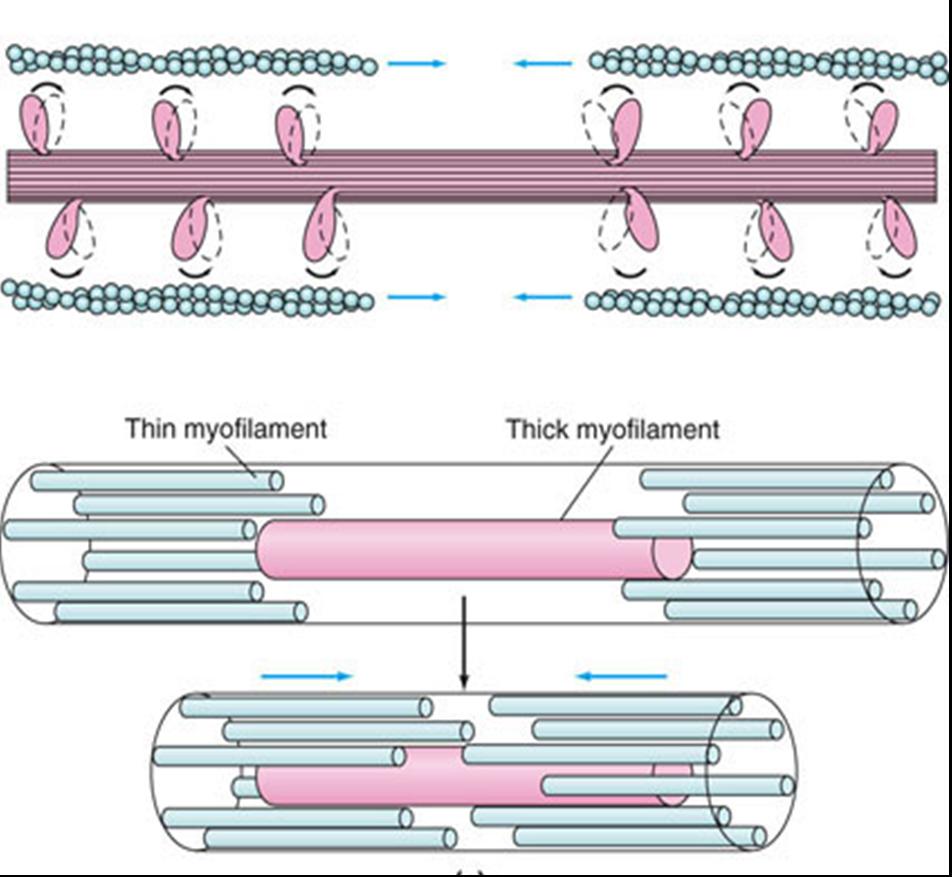
Skeletal Muscle Contraction (Sliding Filament Model)
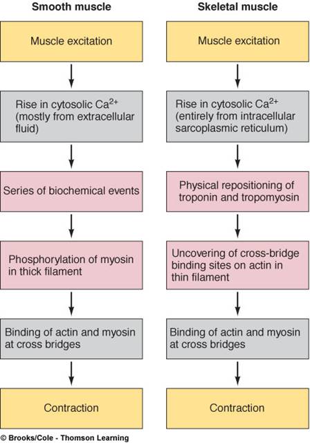
Excitation-Contraction Coupling is the sequence of events linking the transmission of an action potential along the sarcolemma to muscle contraction (the sliding of myofilaments)
The sarcolemma, like other plasma membranes is polarized; there is a potential difference (voltage) across the membrane
Also, ATP attached to the myosin head is split by ATPase causing the myosin heads to be activated.
Steps to Excitation-Contraction Coupling and Muscle Contraction:
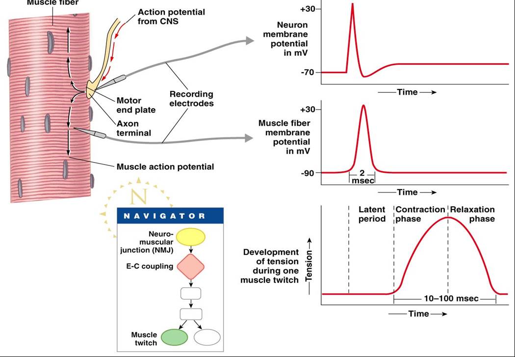
1. The neurotransmitter acetylcholine (ACh) binds to its receptors on the motor end plate
2. Chemically (ligand) gated ion channels in the receptors open and allow Na+ and K+ to move across the membrane, resulting in a transient change in membrane potential (a process called depolarization)
- (End plate potential is a local depolarization that creates and spreads an action potential across the sarcolemma)
- The action potential lasts only 1-2 milliseconds (ms) and ends before contraction occurs
- The period between action potential initiation and the beginning of contraction is called the latent period; excitation-contraction coupling occurs within the latent period.
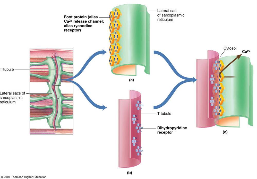
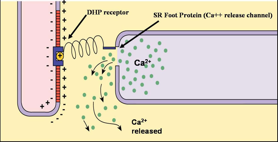
3. The action potential is propagated along (across) the sarcolemma and travels through the T tubules
4. At the triads, the action potential causes voltage sensitive T- tubule dihdropyridine receptors to be activated.
5. The activation of the dihydropyridine receptors causes the sarcoplasmic reticulum foot proteins of the terminal cisternae (lateral sacs) to open Ca2+ release channels
6. Ca2+ is released into the sarcoplasm (where the myofilaments are)
Click here for an animation that describes how an action potential is propagated along the sarcolemma and through the T-tubules and what happens to the calcium ions that are released (next step). This animation is followed by practice questions.
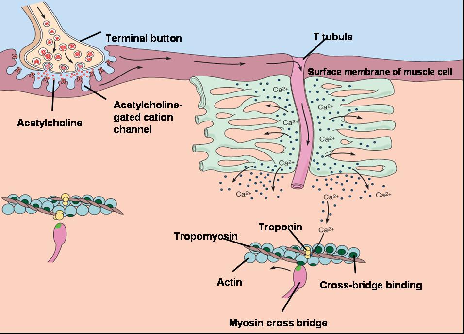
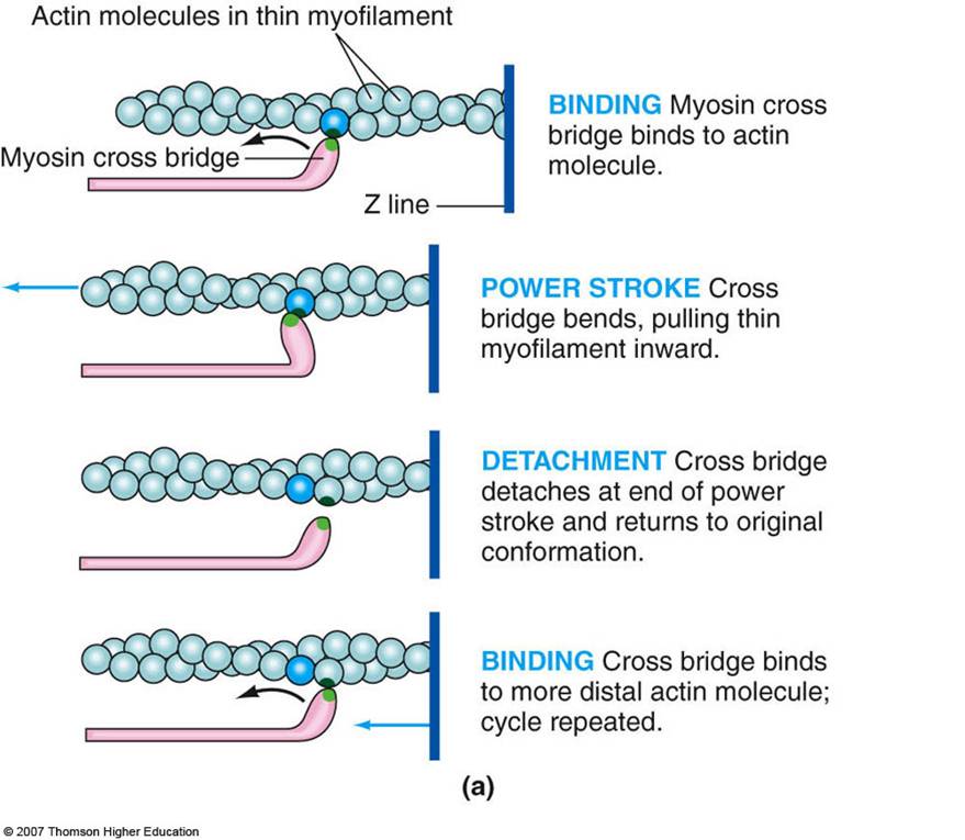
7. The released calcium iona combine with troponin
8. Troponin then pulls the tropomyosin, changing its position so that the actin binding sites are exposed.
Click here for an animation that describes the structure of actin and myosin filaments and how myosin heads pull the actin filaments towards the center of the sarcomere. This animation is followed by practice questions. ( link pending )
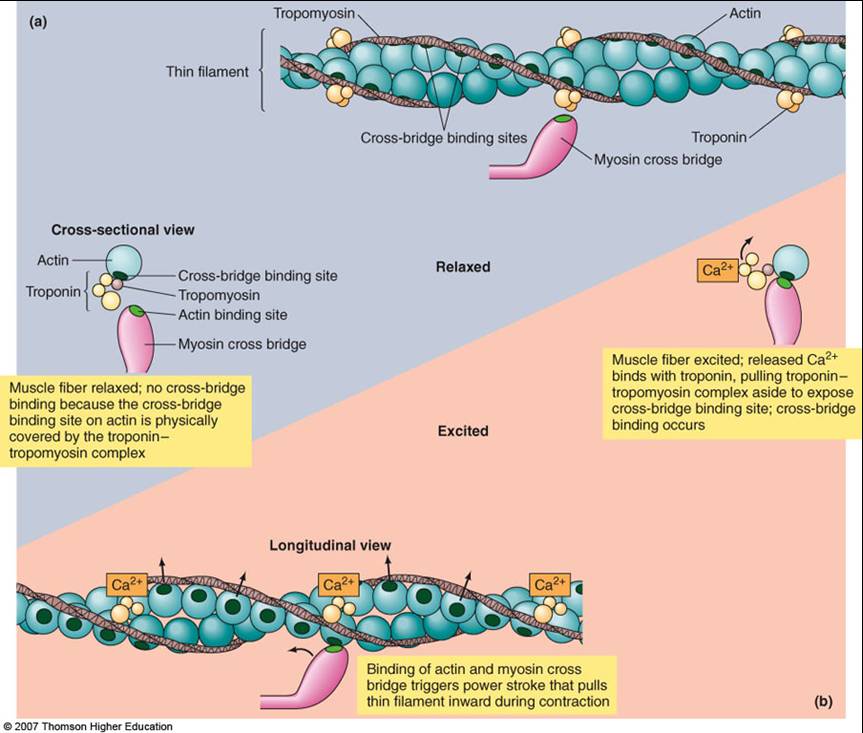
9. The activated myosin head attaches to the actin binding site; contraction refers to the activation of myosin’s cross bridges – the sites that generate the force
10. The phosphate group, P, on the myosin head falls off
11. The myosin head then swivels, producing a power stroke which results in the sliding of the filaments. The ADP on the myosin also falls off.
12. Once the power stroke is complete, ATP again attaches to the myosin head causing the head to detach from the actin site and return to its original position.
13. ATP attached to the myosin head is split by ATPase causing the myosin heads to be activated again; the cycle can then be repeated over and over again as long as calcium and ATP are present.
14. Relaxation is caused by the breaking down of acetylcholine (ACh) by the enzyme acetylcholinesterase and the reabsorption of calcium back into the sarcoplasmic reticulum.
Click here for an animation on muscle contraction. The animation is followed by practice questions.
|
Muscle Tension Physiology
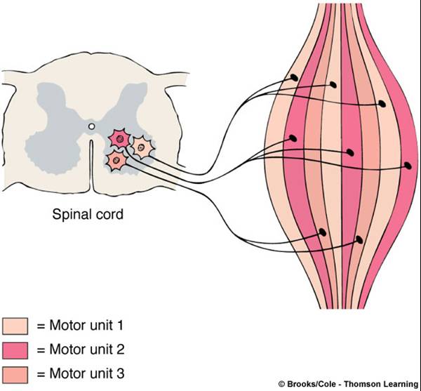
- Motor unit - One motor neuron and the muscle fibers it innervates
- Number of muscle fibers varies among different motor units
- Number of muscle fibers per motor unit and number of motor units per muscle vary widely
- Muscles that produce precise, delicate movements contain fewer fibrs per motor unit
- Muscles performing powerful, coarsely controlled movement have larger number of fibers per motor unit
|
 |
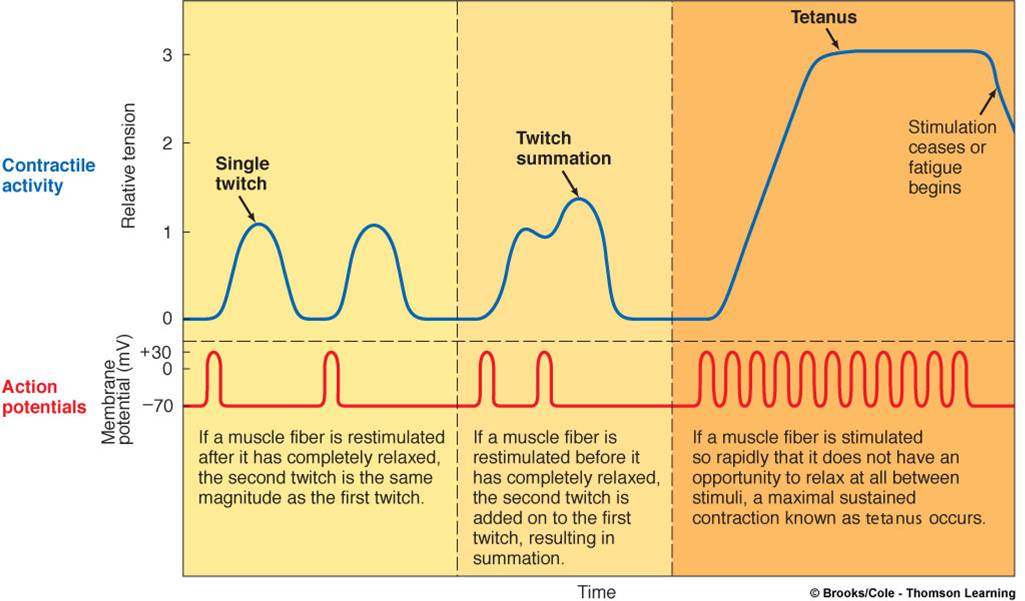
Individual muscle fibers contract to their fullest extent; they do not partially contract, this follows the all or none principle.
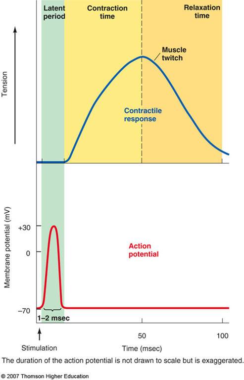
A twitch contraction is a brief contraction of all the muscle fibers in a motor unit in response to a single action potential. It involves 3 phases which can be recorded on a myogram:
- Latent Period
- Contraction Period
- Relaxation Period
|
1. Latent Period
This is the time elapsed from the application of a stimulus to the beginning of the contraction (when calcium ions are being released)
2. Contraction Period
Cross bridges are active and the muscle shortens if the tension is great enough to overcome the load
3. Relaxation Period
Active transport of Ca++ back into the sarcoplasmic reticulum and degradation of acetylcholine
|
 |
Graded Muscle Responses
- These are variations in the degree or strength of muscle contraction in response to demand, required for proper control of skeletal movement
- Muscle contraction can be graded (varied) in two ways:
- Frequency of the stimulation
- Number of motor units stimulated
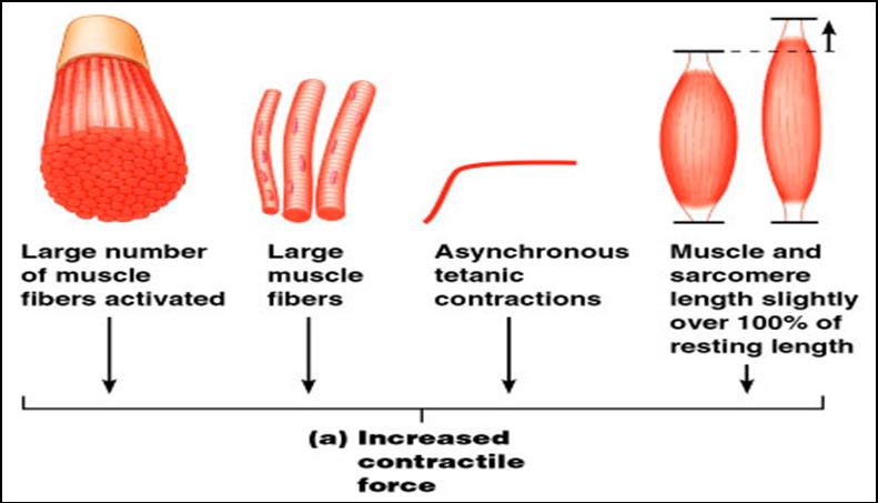
Motor Unit Recruitment
- This is the process of increasing the number of active motor units in a muscle for stronger contractions
- A single stimulus results in a single contractile response – a muscle twitch (contracts and relaxes)
- More frequent stimuli increases contractile force – wave summation - muscle is already partially contracted when next stimulus arrives and contractions are summed

- A sustained contraction that lacks even partial relaxation is known as tetanus.
- Asynchronous recruitment of motor units is used to prevent fatigue. While some motor units are active others are inactive. This pattern of firing of motor neurons prevents fatigue while maintaining contraction by allowing a brief rest for the inactive units.
Recruitment also helps provide smooth muscle action rather than jerky movements.
Muscle Tone
- The constant, slightly contracted state of all muscles
- Does not produce active movements
- Keeps the muscles firm and ready to respond to stimulus
- Helps stabilize joints and maintain posture
- Due to spinal reflex activation of motor units in response to stretch receptors in muscles and tendons
Strength of Muscle Tension
Muscle tension is the force exerted on an object by a contracting muscle
- Muscle tension depends on:
- Number of motor units recruited
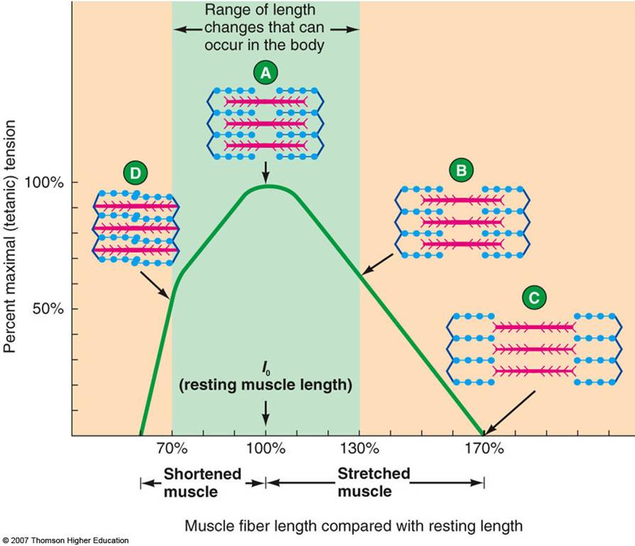
- Degree of muscle stretch
- Relative size of the muscle fibers – the bulkier the muscle fiber (greater cross-sectional area), the greater its strength
- Recruitment also helps provide smooth muscle action rather than jerky movements
- Asynchronous recruitment of motor units provides a brief rest for the inactive units preventing fatigue; in this pattern of firing, some motor units are active others are inactive .
Types of Contractions
- Muscle tension is the force exerted on an object by a contracting muscle
- Load is the opposing force or weight of the object to be moved
- There are 2 types of muscle contractions:
1. Isometric
- The muscle does not or cannot shorten, but the tension on the muscle increases

2. Isotonic
- Moving a constant load through the range of muscle motion
- There are 2 types of isotonic contractions:
1. Concentric - the muscle shortens and pulls to produce a movement to reduce the angle at a joint
2. Eccentric - the overall muscle lengthens during a contraction

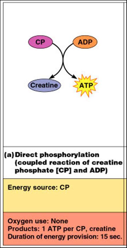
Metabolism
ATP is the only energy source that is used directly for contractile activity
Several pathways supply ATP to muscle cells:
- Transfer of high-energy phosphate from creatine phosphate (CP) to ADP
- Oxidative phosphorylation
- Glycolysis
|
1. Transfer of high-energy phosphate from creatine phosphate (CP) to ADP
- First energy storehouse tapped at onset of contractile activity.
- Transfer of energy as a phosphate group is moved from CP to ADP
- the reaction is catalyzed by the enzyme creatine kinase
- Creatine phosphate + ADP → creatine + ATP
- Stored ATP and CP provide energy for maximum muscle power for 10-15 seconds
|
 |
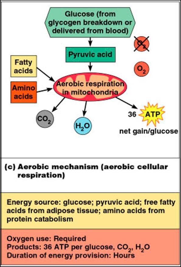
2. Oxidative Phosphorylation (citric acid cycle and electron transport system)
- This takes place within muscle mitochondria if sufficient O2 is present
- Glucose is broken down into pyruvic acide to yield 2 ATP
- When oxygen demand cannot be met, pyruvic acid is converted into lactic acid
- Lactic acid diffuses into the bloodstream – can be used as energy source by the liver, kidneys, and heart
- Can be converted back into pyruvic acid, glucose, or glycogen by the liver
|
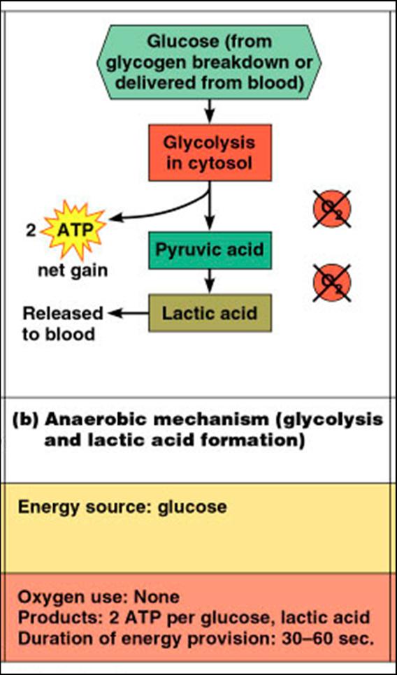 |
3. Glycolysis
- Is a series of reactions breaks down glucose for high yield of ATP
- Supports anaerobic or high-intensity exercise,
Aerobic respiration occurs in mitochondria -
- It requires O2
- Glucose + O2 → CO2 + H2O + ATP
|
 |
Muscle Fatigue
- The muscle is physiologically not able to contract
- Occurs when oxygen is limited and ATP production fails to keep pace with ATP use
- Lactic acid accumulation and ionic imbalances may also contribute to muscle fatigue
- Depletion of energy stores – glycogen
- When no ATP is available, contractures (continuous contraction) may result because cross bridges are unable to detach
- Insufficient O2,
- Build up of lactic acid
Depletion of energy reserves - glycogen
Ionic imbalance
- Neural fatigue
- Central Fatigue – psychological, it hurts
For a muscle to return to its pre-exercise state:
- Oxygen reserves must be replenished
- (Lactic acid must be converted to pyruvic acid)
- Glycogen stores must be replaced
- ATP and CP reserves must be resynthesized
Oxygen Debt
This is t he extra amount of O2 needed for the above restorative processes |
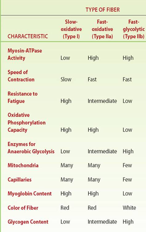
Muscle Fiber Types
Type I Slow Oxidative - fatigue resistant fibers containing large amounts of myoglobin, many mitochondria, a many capillaries, high capacity for generating ATP, slow contraction rate. Abundant in postural muscle groups
Type IIA Fast Oxidative - same as Type I except fast contraction rate. Abundant is muscle groups requiring speed (sprinter)
Type IIB Fast Glycolitic - easily fatigueable fibers, low myoglobin, few mitochondria, few capillaries, large amounts of glycogen , large diameter fibers used in muscles requiring strong and rapid, but brief contractions (arms).
- Speed of contraction – determined by how fast their myosin ATPases split ATP
- Oxidative fibers – use aerobic pathways
- Glycolytic fibers – use anaerobic glycolysis
|
 |
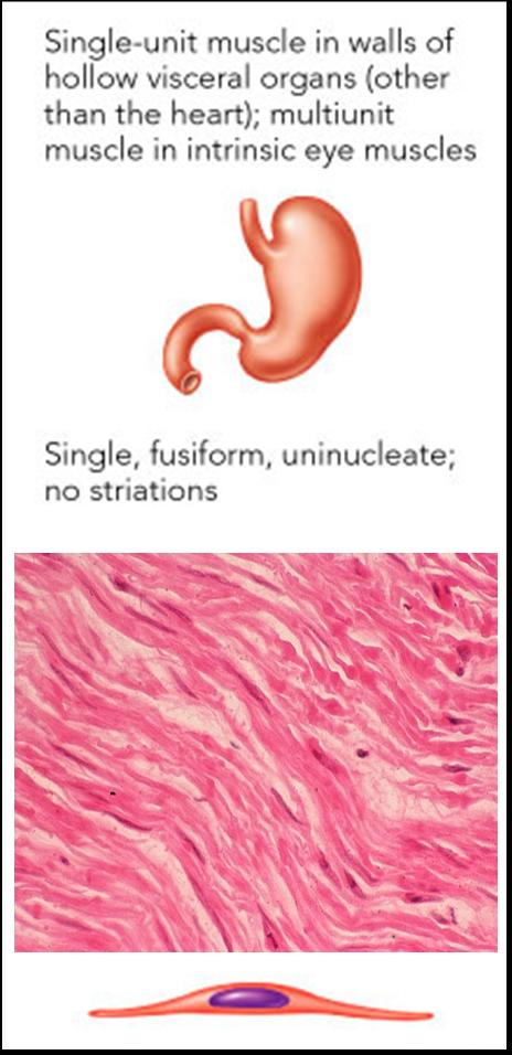
Smooth Muscle
- Occurs within most organs
- Walls of hollow visceral organs, such as the stomach
- Urinary bladder
- Respiratory passages
- Arteries and veins
- Helps substances move through internal body channels via peristalsis
- No striations
- Filaments do not form myofibrils
- Not arranged in sarcomere pattern found in skeletal muscle
- Is under involuntary control
- Single nucleus within each muscle cell (muscle fiber)
- Composed of spindle-shaped fibers with a diameter of 2-10 mm and lengths of several hundred mm
|
 |
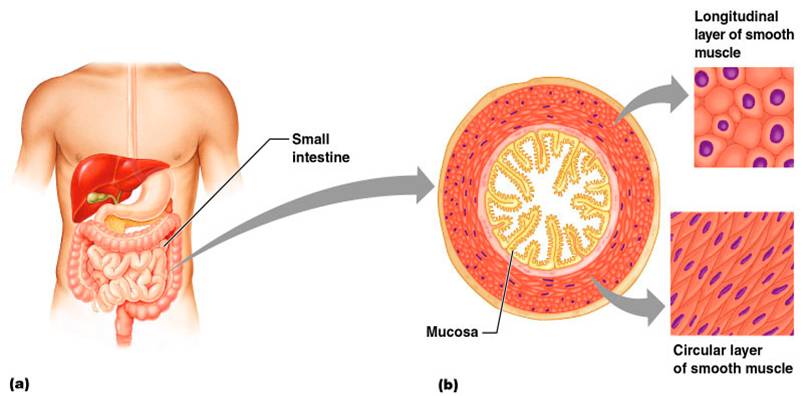
Smooth Muscle Contraction
- Cells usually arranged in sheets within muscle
- Organized into two layers (longitudinal and circular) of closely apposed fibers
- Have essentially the same contractile mechanisms as skeletal muscle
|
- Cell has three types of filaments
- Thick myosin filaments
- Longer than those in skeletal muscle
- Thin actin filaments
- Contain tropomyosin but lack troponin
- Filaments of intermediate size
- Do not directly participate in contraction
- Form part of cytoskeletal framework that supports cell shape
- Have dense bodies containing same protein found in Z lines
|
 |
- Whole sheets of smooth muscle exhibit slow, synchronized contraction
- Smooth muscle lacks neuromuscular junctions
- Action potentials are transmitted from cell to cell
- Some smooth muscle cells:
- Act as pacemakers and set the contractile pace for whole sheets of muscle
- Are self-excitatory and depolarize without external stimuli
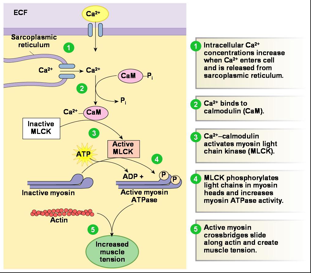
- Muscle fiber stimulated
- Ca2+ released into the cytoplasm from ECF
- Ca2+ binds with calmodulin
- Ca2+/Calmodulin activates mysoin kinase
- Myosin kinase phosphorylates myosin
- Myosin can now bind with actin
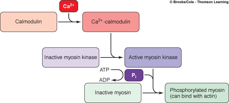
Ca2+ for smooth muscle contraction comes largely from outside the cell.
|
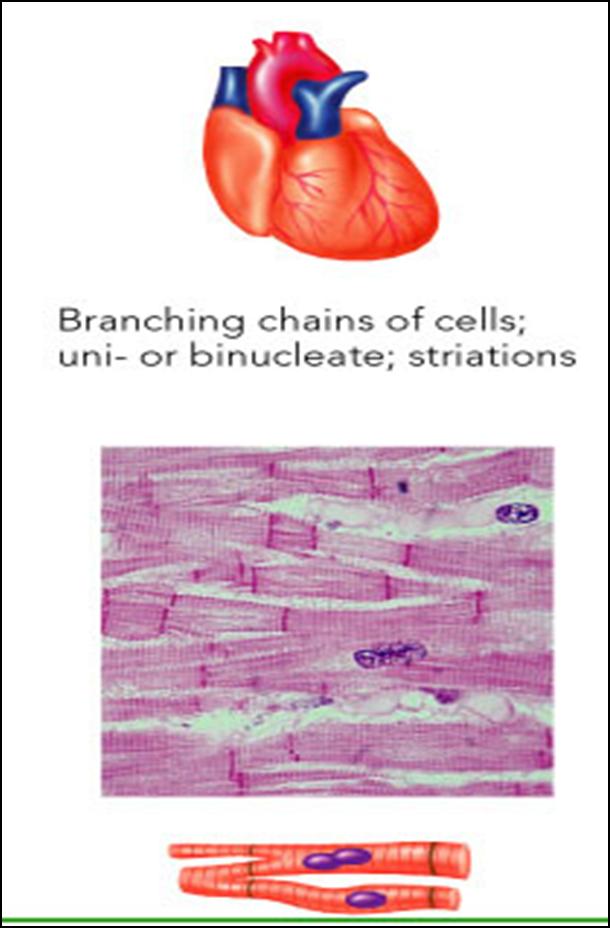
Cardiac Muscle
- Occurs only in the heart
- Is striated like skeletal muscle but but has a branching pattern with intercalated discs
- Usually one-two nuclei per muscle cell
- Is under involuntary control
- Contracts at a fairly steady rate set by the heart’s pacemaker
- Neural controls allow the heart to respond to changes in bodily needs
|
 |
|