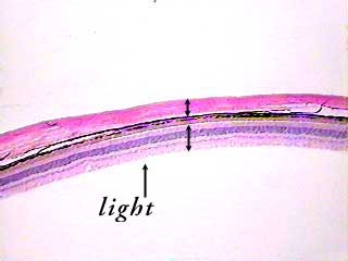
This image shows a cross section of part of the wall of the eyeball.
The eyeball has three layers: the outer fibrous tunic, the middle
vascular tunic, and the inner sensory tunic. The part of the
fibrous tunic shown here is the sclera (top arrow bar). Just
below it, but not labeled, is the choroid (part of the vascular
tunic). On the bottom of the image is the retina, or sensory
tunic (bottom arrow bar). You can see even at this low magnification
that the retina is made of several layers.
This section is from somewhere in the back of the eye. Light would enter the eye and move through several other structures before reaching the retina. The arrow shows the direction of light movement through the eye.
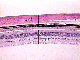
The sclera (scl) is made of dense, avascular connective tissue.
The choroid is made of loose connective tissue. Its outer layer contains a large amount of the brown pigment melanin (looks like a black stripe in this image). The inner layer of the choroid is not pigmented. Here it looks like a pink stripe underneath the black stripe.
The innermost layer of the eyeball, the retina, is composed
of three layers of nerve cells that will be easier to see in
the next image.
Compare this to the photomicrographs in your textbook. They may
not show the retina at the same magnification, and they may not
show all of the layers.
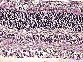
This image shows only the retina, which has two parts. On the very top of this image, you can see the pigmented epithelium of the retina where it is separated from the choroid. (The pigmented epithelium of the retina is not the same pigmented layer that is visible as a black stripe in the other images on this page.) The retinal pigmented epithelium is simple columnar, so it is very thin. In this image, it is not very dark, but you can see a brownish line at the top. The rest of the image shows the nervous layer of the retina. It has three main layers: on top are the photoreceptors--rods and cones. The actual rod and cone part of the cell is in the layer labeled r/c. The layer just below that contains the cell bodies of the photoreceptors. The densely packed nuclei make this layer look very dark, but it is not pigmented. The layer labeled b contains the nuclei of the bipolar cells. The layer on the bottom, labeled g, contains the nuclei of the ganglion cells.