Parotid Salivary Gland, 40X

 
There are nine images on this page--three magnifications each of the three types of salivary glands (parotid, submandibular, 
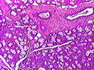
and sublingual. The first thing you have to know about the salivary 
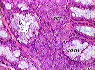
glands is the difference between serous cells and mucous cells.

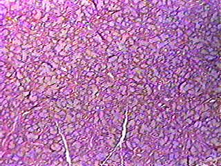
Mucous cells produce mucus, which doesn't stain very darkly, so the mucous cells look almost clear on these images and on slides. Serous cells produce a watery secretion that contains a lot of proteins. Serous cells stain fairly dark. Each of the three glands has different proportions of these two cell types. The parotid glands contain nothing but serous cells. The submandibular glands contain both mucous and serous cells. The sublingual glands contain mostly mucous cells with just a few serous cells. You can use this information to figure out which of the three salivary glands you are looking at.

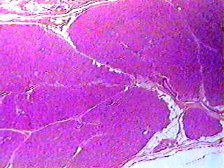
The real problem is that the parotid gland looks a lot like the pancreas. Both of them usually stain the same color and most of the cells are arranged in the same pattern. But the pancreas contains small clumps of cells called pancreatic islets or islets of Langerhans that stain lighter and have a different pattern of arrangement than the other part of the pancreas. So, you will know you are looking at the pancreas when you do see the islets, and the parotid salivary gland when you don't see the islets.


Parotid Salivary Gland, 100X

 
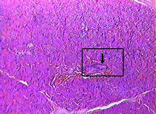
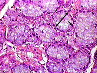
You can see ducts and blood vessels on most slides of salivary glands. The arrow in the box points to a cross section of a duct. Ducts are easy to recognize because most of them are lined by simple cuboidal epithelium. Larger ducts may have simple columnar, stratified cuboidal or even stratified columnar epithelium.

Just below the duct and to its left is a blood vessel--it looks more red than the surrounding tissue due to the presence of red blood cells.


Parotid Salivary Gland, 400X
 
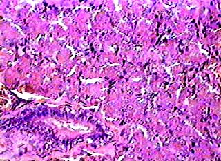
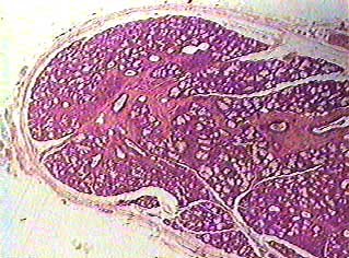
This is an enlargement of the area in the box in the image above. You can see the duct in the lower left corner, and part of a blood vessel just above it.


Sublingual Salivary Gland, 40X
 
The light-staining spots all over this image are clumps of mucous cells. The mucus accumulates in the apical (surface side) of the cell, and the nucleus and cytoplasm are pushed towards the bottom (basal side) of the cell. The part of the cells where mucus is found are light in color because mucus does not accept the kind of stain used on this slide very well. The dark ring you see surrounding the light areas is caused by the nuclei, which are usually darkly stained.

Sublingual Salivary Gland, 100X
 
Note again the mucus secretory units that appear light in the center with a dark ring around the outside. Do you see any ducts?


Sublingual Salivary Gland, 400X
 
The arrow bar indicates the diameter of one mucous secretory unit. Note again the location of mucus and nucleus (small arrow) in the individual cells. What kind of epithelium lines the duct in the lower left part of the image?


Submandibular Salivary Gland, 40X
 
The submandibular gland should look to you like a cross between the other two salivary glands. You can see the light spots that represent mucous cells, but there are a lot of dark areas representing clumps of serous cells as well.


Submandibular Salivary Gland, 100X
 
At the top of this image are cross sections of several ducts that are located in the septa ("walls" of connective tissue that separate the salivary gland into lobes). They are larger than the ducts found in the secretory parts of the gland and may be lined with stratified cuboidal epithelium.


Submandibular Salivary Gland, 400X
 
One serous-secreting unit (ser) and one mucus-secreting unit are labeled on this image. Serous cells are usually arranged in spherical bundles that secrete into a duct. Mucous cells are often arranged in tubes, not spheres. The mucus is secreted right into the tube, which is also the duct for those cells.


return to organ slide index