Duodenum - 40X
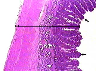
The wall of the duodenum has the same four layers as the wall of the stomach.
The position and relative thickness of each layer is shown on the bar. From
right to left the layers are: mucosa, submucosa, muscularis and serosa.
You can see that the muscularis is further subdivided into two layers of
smooth muscle.
The arrows point to villi--microscopic finger-like projections of the
mucosa. Look carefully at the villi--only the epithelium and lamina propria
are involved. The muscularis mucosae does not project up into the villi.
Duodenum - 100X
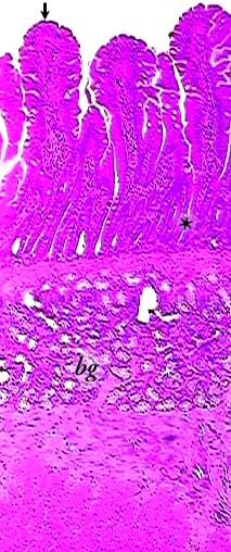
Although it is not very clear on this image, between the bases of the villi are tubular glands called intestinal crypts (*). The openings of these glands are between the bottoms of the villi.
In the submucosa of the duodenum are the duodenal or Brunner's glands (bg) that secrete alkaline mucus.
Below the Brunner's glands are the smooth muscle layers of the muscularis.
Duodenum - 400X
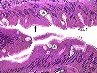
The light stripe (arrow) that seems to run along the surface of the simple columnar epithelium is caused by the presence of microvilli on the surface of the epithelial cells. This is sometimes called the "brush border" because (at higher magnification) they look like the bristles of a brush.
Some of the light circular areas in the epithelium (*) are mucus in goblet cells.