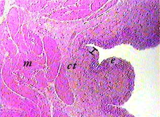
The wall of the urinary bladder has four layers. From the inside
towards the outside they are: mucosa, submucosa, muscularis,
and serosa or adventitia. The outside layer is either serosa
or adventitia, depending on location--see your textbook for an
explanation. Some authorities do not include a submucosa in the
wall of the urinary bladder--in their opinions, all of the connective
tissue (ct) between the mucosa and the muscularis is part of
the lamina propria of the mucosa.
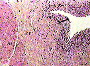
In this image, the thickness of the epithelial part of the
mucosa--transitional epithelium-- is shown by the bar and labeled
e. To the left of the epithelium is the connective
tissue (ct) that includes both the lamina propria of the mucosa
and the submucosa. To the left of the connective tissue is the
muscularis of the urinary bladder, which is called the detrussor
muscle.
The bar shows the thickness of the transitional epithelium lining the urinary bladder. To the left of the epithelium is connective tissue comprising the lamina propria of the mucosa and the submucosa. To the right of that is smooth muscle of the muscularis.
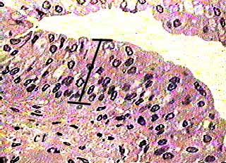
If you can recognize this as transitional epithelium, you can narrow the location down considerably. It is found in only three organs: ureters, urinary bladder, and part of the urethra. The ureter and urethra are tubes and would probably look like a cross section of circular structure with a lumen. On the other hand, a slide of the urinary bladder can include only a portion of the organ's wall.