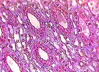
Kidney cortex
This is the same image we used to show you how to find simple
squamous epithelium in the kidney (the outer part of the kidney
is at the top). Since each organ in the body is made of two or
more tissues, most of the slides you see in lab will have several
tissues on them. So you have to learn 1) where to find the tissue
you want and 2) how to tell it apart from all of the other tissues
that are one the same slide.
This time you are looking for simple cuboidal epithelium. It is found throughout the kidney. It forms most of the microscopic tubes that process body fluids and make urine. You can find it near the glomeruli (round structures in top third of image) and also in the lower parts of the kidney
(The bar will be explained later.)
Kidney cortex
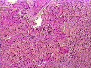
Cuboidal cells are about as tall as they are wide. The nucleus
is usually round and located in the center of the cell. Simple
epithelial tissues have only one layer of cells.
Knowing about the shape and position of the nucleus in the
cells will allow you to recognize tissues even when you can't
tell where the edges of the cells are. Since you are trying to
find a simple cuboidal epithelium, look for rows or rings of
round dark dots (the nuclei of the cuboidal epithelial cells).
Most of the cells in this image are simple cuboidal epithelial
cells.
Kidney cortex
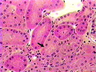
The arrow is pointing to a cross section of a tube made of simple
cuboidal epithelium. Start at the point of the arrow and follow
the nuclei in a circle until you get back to the arrow. There
are several other tubes shown in cross section on this image.
Some of the tubes were sliced (sectioned) straight across
and they look like circles. In other places the tubes were cut
at an angle (in anatomy this is called an oblique section) and
they look more like ovals. In the inner parts of the kidney (on
the middle third of the top image on this page) the tubes are
sliced longitudinally (long wise) and the cells that make up
the walls of the tubes appear in parallel rows.
Kidney medulla
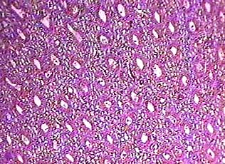
This image is from the part of the kidney indicated by a bar
in the top image on this page. You are now looking at the same
structures but from a different angle. All of the structures
in this image are sections of kidney tubules.
Kidney medulla
This image is an enlargement of the one just above it. The color of the stain is different from the top three images, but you can still recognize the pattern formed by the nuclei of the simple cuboidal epithelial cells--just look for the circles. The individual cells can be seen in this image, making the simple cuboidal epithelium even easier to recognize.