Human respiratory tract
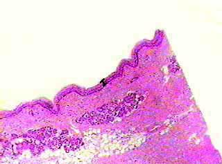
The bar in this image shows the thickness of the layer of ciliated pseudostratified epithelium. The rest of the tissue below the epithelium is mostly connective tissue, but there are some mucous glands (glandular epithelium) just below the surface. At this magnification you should practice locating the epithelial layer, which is lining part of the respiratory tract. Epithelial tissues that form coverings or linings always have one free (unattached) surface.
Human respiratory tract
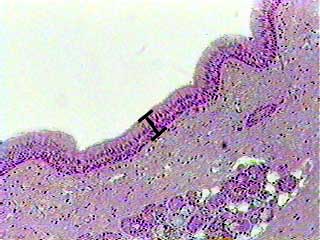
At 100X you can begin to seee the individual cells that make up pseudostratified epithelium. Notice that most of the cell nuclei tend to be along the bottom (basal) edge of the tissue.
Human respiratory tract
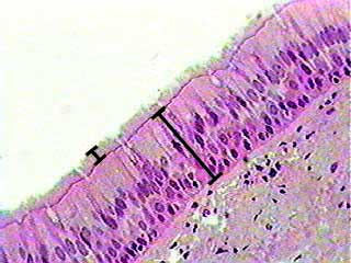
Now you can see the individual cells. Once again, the bar shows you the thickness of the ciliated pseudostratified epithelium. The cells in this tissue are very tall and thin, and are not all the same shape. All of the cells are in contact with the basement membrane (on the basal surface), but not all of them reach the surface. The cells that do reach the surface are either ciliated or goblet cells (mucus-secreting cells). The cells that do not reach the surface are probably stem cells and will eventually replace the ciliated and goblet cells. The small bar shows the location and height of the cilia.