(Palmar skin)
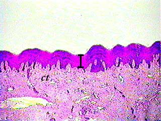
Although stratified squamous keratinized epithelium covers the entire surface of the body, most of it also includes hair, which makes the basic tissue structure harder to see. If we just want to look at stratified squamous keratinized epithelium, we look at skin from one of the few areas of the body that does not have hair. This tissue is from the palm of the hand (palmar skin). The bar shows the thickness of the stratified squamous keratinized epithelium. In this specimen, the epithelium is stained very dark. The lighter areas underneath are connective tissue (ct).
(Palmar skin)
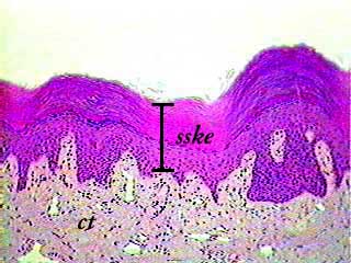
At 100X you can see the distinct cell layers that make this a stratified epithelium. The bar indicates the thickness of the stratified squamous keratinized epithelium (sske). Notice that the nuclei of the cells in the bottom layers tend to have a round shape, but that the nuclei seem to become flatter as you move towards the surface. You have probably noticed that the bottom surface of the epithelium is not flat. The bumpy appearance is caused by connective tissue projections called dermal papillae. On all of the images on this page, the epithelium is stained more darkly than the connective tissue.
(Palmar skin)
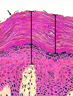
The cells on the surface of stratified squamous keratinized epithelium are very flat. Not only are they flat, but they are no longer alive. They have no nucleus or organelles. They are filled with a protein called keratin, which is what makes our skin waterproof. The arrow at the top of the image is pointing to a keratinized cell that has partially separated from the rest of the skin. These dead cells are continually lost from the surface of the skin, and are replaced by new cells from the layers below. The cells on the basal (bottom) layer are actually cuboidal or columnar in shape. They divide by mitosis to produce a constant supply of new cells that replace the ones that are lost from the surface. As these replacement cells are gradually pushed towards the surface, their shape changes. They start out on the bottom as cuboidal cells, then they become irregular in shape, and finally become very flat as they are transformed in the dead, keratin-filled surface cells.