Silver stain
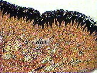
Dense irregular connective tissue (dict) is found in several places in the body. This image is from palmar skin (skin from the palm of the hand), and the dense irregular connective tissue is stained light brown. The very dark tissue on the top of the image is stratified squamous keratinized epithelium.
The reason the colors look so strange in the images on this page is the silver stain that was used to emphasize the location of collagen and reticular fibers. You will also see the more traditional hematoxylin (blue) and eosin (pink) on other skin slides (stratified squamous keratinized epithelium). You have to learn to recognize the tissue from its location relative to other structures and by any patterns that you can see in the cells or extracellular materials. Learning to recognize a tissue by the color it was stained will get you into trouble on a practical exam if the instructor uses a different slide.
Silver stain
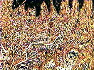
Most of the tissue you see in this image is dense irregular connective tissue (dict). There is a small amount of epithelium on the top (black) and some adipose tissue at the bottom and lower left (very light). Most of the rest of the image shows bundles of collagen fibers, which are stained brown.
Silver stain
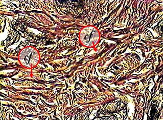
In this image you can see the collagen fibers (cf) that are the main component of dense irregular connective tissue. The n fibroblasts that make the collagen fibers cannot be seen because they do not pick up as much of the stain as the collagen fibers do.