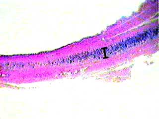
Cartilage is easy to recognize because it looks so much different from other tissues. This image shows a section of the wall of the trachea. You can feel the hyaline cartilage in your own trachea by pressing you fingers gently against the front of your throat and moving them slightly up and down. The hyaline cartilage in the trachea is in the middle of the tracheal wall. It tends to stain more blue than other kinds of connective tissue (however, remember that color should never be the main cue you use to identify a tissue). The bar shows the position of the hyaline cartilage.
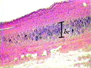
You can begin to see the details in hyaline cartilage (hc) structure in this image. The bar shows you the extent of the cartilage in the tracheal wall. At the very top of this image is a layer of pseudostratified ciliated epithelium. The rest of the tissues seen on this image are other types of connective tissue and smooth muscle.

Cartilage consists of cells embedded in a matrix (mat) of fibers and ground substance. The cells are called chondrocytes (ch) and the spaces in the cartilage in which they are found are called lacunae. Hyaline cartilage has very few fibers in its matrix, so the matrix usually looks smooth. The cells you see in the upper left corner of this image are part of the perichondrium, which consists of dense connective tissue.