40X
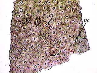
The images on this page are made from bone that still has the calcium salts in its matrix. The difference is that it is ground a lot thinner than the bone specimen on the previous page, so we can see more detail. You can see many osteons on this image. Each one has a light spot in the center where the Haversian or central canal (cc) is located. Another type of canal--perforating or Volkmann's (vc)--connect the central canals to each other. Not every slide you see in lab will show the Volkmann's canals, so you may have to look at 2 or 3 different slides until you find one.
100X
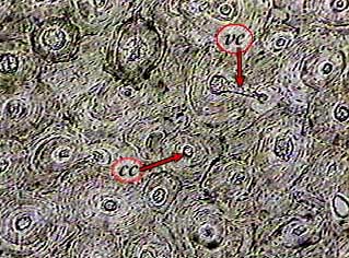
Haversian or central canals (cc) are found in the center of an osteon, but perforating or Volkmann's canals (vc) cut across the osteons to connect two central canals.
400X
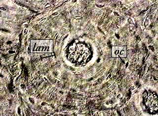
A lamina (lam) is a layer of matrix between two concentric "rows" of osteocytes. It is difficult to know where one lamina begins and ends, because the osteocytes are not arranged in neat rows. The number of rows is limited by the distance nutrients can diffuse from the blood vessel in the central canal to the osteocytes. The outermost osteocytes can't be more than about 0.2 mm from the central canal.