Skeletal muscle can be confused with dense regular connective
tissue at low magnification (especially 40X). They stain the
same color, and the skeletal muscle cell nuclei are flattened
just like the fibroblast nuclei in dense regular connective tissue.
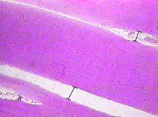
In this image you are looking at three bundles of skeletal muscle cells (fascicles). The bars show you the location of the connective tissue (perimysium) that separates the bundles.
Some of the purple dots you see in the image are the nuclei
of the skeletal muscle cells, but some of the purple dots are
artifacts of the digitizing procedure.
Although the resolution of this image does not reveal the edges
of individual muscle cells, you can tell from the position of
the nuclei where the cells are located. The nuclei are pushed
to the edge of the cell by the proteins that allow the cell to
contract. Where you see lines of flattened purple dots, you are
looking at one side of a muscle cell.
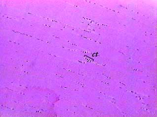
There is connective tissue (endomysium) between the muscle
cells. The nuclei of the connective tissue cells (fibroblasts)
in the connective tissue (ct) may be smaller and rounder than
the nuclei of the skeletal muscle cells.
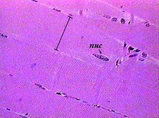
The bar shows the width of one skeletal muscle cell. Most of
the muscle cell nuclei you see will be along the sides of the
cells. The nucleus identified in the image (nuc) is just inside
the cell membrane, but the top the cell was caught by chance
in this section. When you look at skeletal muscle cells sectioned
longitudinally the nuclei will look long and flat or oval. When
you look at cells that were sectioned transversely (cross section)
the nuclei will look like round dots. The images on this page
only show cells that are sectioned longitudinally.
The faint lines that run across the cells are called striations.
They are not actual structures inside the cell, but are caused
by the way the light from the microscope shines through the proteins
inside the cell. Because the proteins are lined up precisely,
they scatter the light as it passes through the specimen and
makes a striped or banded pattern. If you cannot see the striations
in lab, try closing the iris diaphragm a little to increase contrast,
and then use the fine focus knob to focus up and down until the
striations appear.