Smooth muscle can be confused with cardiac muscle because the
cells are often running in different directions, just as they
are in cardiac muscle. Smooth muscle cells are a lot smaller
than cardiac muscle cells, and they do not branch or connect
end to end the way cardiac cells do.
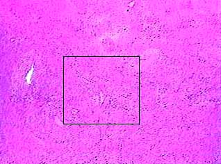
The area inside the box is enlarged in the next image.
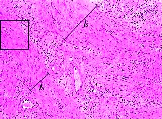
To get an idea of the arrangement of the individual cells, look at the nuclei, which look like purple spots in this image. If the nuclei look long and thin, the cells have been sectioned longitudinally (ls). If the cells look round, the cells have been sectioned transversely.
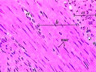
This image provides an even better comparison of smooth muscle cells that have been sectioned in different planes (ls and cs). The nuclei (nuc) of smooth muscle cells are located in the center of the cell. Even though you can't see the cell membranes or the edges of the cells, you can visualize their arrangement just by looking at the nuclei.