Motor Neuron smear
There are many different kinds of cells in the nervous system,
but they can be organized into two major categories: neurons
and supporting cells. Neurons (n) are the ones that generate
and conduct nerve impulses. Supporting cells do not conduct nerve
impulses, but they perform many other functions for the nerve
tissue.
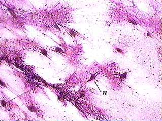
The images on this page were made from a slide called a motor
neuron smear. Motor neurons are large and easy to see, so they
are usually used as examples. A smear means that a small chunk
of nerve tissue from the spinal cord or brain was literally squashed
and spread out on a slide. That's the only way to see neurons,
because they have many extensions that would be cut off in a
typical section.
Motor Neuron smear

Each neuron (n) has extensions called processes (axons and dendrites) that allow it to communicate with other neurons. The pink lines that are attached to these neurons are their processes. About 90% of the cells in the central nervous system--brain and spinal cord--are supporting cells (sc). You can see that they are much smaller than neurons.
Motor Neuron smear
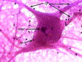
Do you recognize this image? Tilt your head to the right and look again. We used this neuron as our mascot on the main page. The small dark dots are probably all nuclei of supporting cells that are either on top of or underneath the neuron. The large dark spot in the neuron is where its nucleus is located. Several cell processes (cp) extend outwards from the main body of the neuron. If you look at the processes where they are attached to the neuron's body, you can see neurofilaments (refer to your text book for an explanation of neurofilaments).