|
|||||
|
|
|
|
|
|
|
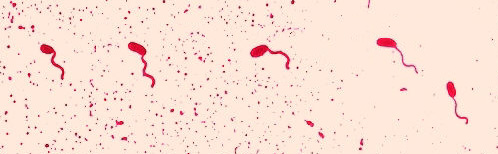
Flagella Stain
The flagella stain allows observation of bacterial flagella under the light microscope. Bacterial flagella are normally too thin to be seen under such conditions. The flagella stains employs a mordant to coat the flagella with stain until they are thick enough to be seen. These staining techniques are typically very difficult. Because of this, you will not be performing a flagella stain this semester, but there are prepared slide of flagella stains available for you to view.
Many bacteria are motile; some accomplish this motility by means of
flagella. Flagella can vary by number and location. Some bacteria
only have one flagella; this is called monotrichous. In most monotrichous
bacteria, the flagella is at the end of the cell; this placement is called
polar. Some bacteria have a flagella at either end of the cell; this
arrangement is called amphitrichous. Many bacteria have multiple flagella;
these may all be located in a tuft at one end of the cell, in which case
the arrangement is lophotrichous, or they may be all over the cell, in
which case the arrangement is peritrichous.
The staining procedure is here.