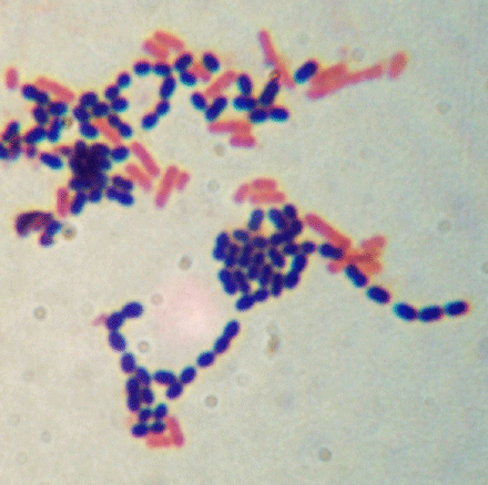
Gram Stain
The Gram stain is the most important staining procedure in microbiology. It is used to differentiate between gram positive organisms and gram negative organisms. Hence, it is a differential stain.
Gram staining involves a four-part process, which includes:
crystal violet, the primary stain
iodine, the mordant
a decolorizer made of acetone and alcohol
safranin, the counterstain
Gram negative and gram positive organisms are distinguished from each other by differences in their cell walls. These differences affect many aspects of the cell, including the way the cell takes up and retains stains.
Gram positive cells take up the crystal violet, which is then fixed in the cell with the iodine mordant. This forms a crystal-violet iodine complex which remains in the cell even after decolorizing. It is thought that this happens because the cell walls of gram positive organisms include a thick layer of protein-sugar complexes called peptidoglycans. This layer makes up 60-90% of the gram positive cell wall. Decolorizing the cell causes this thick cell wall to dehydrate and shrink, which closes the pores in the cell wall and prevents the stain from exiting the cell. At the end of the gram staining procedure, gram positive cells will be stained a purplish-blue color.
Gram negative cells also take up crystal violet, and the iodine forms a crystal violet-iodine complex in the cells as it did in the gram positive cells. However, the cell walls of gram negative organisms do not retain this complex when decolorized. Peptidoglycans are present in the cell walls of gram negative organisms, but they only comprise 10-20% of the cell wall. Gram negative cells also have an outer layer which gram positive organisms do not have; this layer is made up of lipids, polysaccharides, and proteins. Exposing gram negative cells to the decolorizer dissolves the lipids in the cell walls, which allows the crystal violet-iodine complex to leach out of the cells. This allows the cells to subsequently be stained with safranin. At the end of the gram staining procedure, gram negative cells will be stained a reddish-pink color.
Remember:
If staining from a broth, always vortex the broth first.
Only perform a gram stain on cultures that are 24 hours old.
Do NOT decolorize for a full minute! Flood the slide with decolorizer, put the cap back on the bottle, and then rinse! The decolorizer should stay on the slide for no more than 15 seconds! If the decolorizer is left on too long, even gram positive cells will lose the crystal violet and will stain red.
The staining procedure is here.

